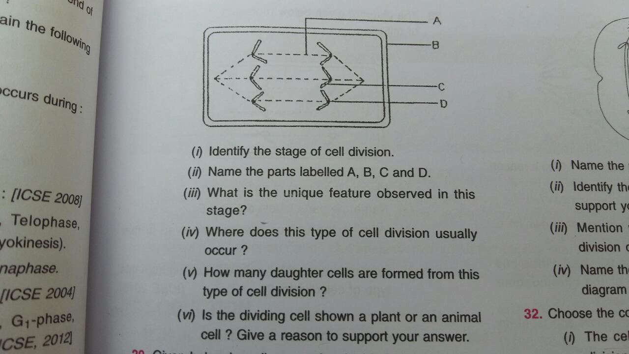
43 label the following diagram of mitosis of an animal cell
Label the following Diagram of mitosis of an animal cell Step 5: Telophase (Two nuclei produced and cell pinches in the middle) Question # 2: Interphase . Question # 3: Centrioles are the organelles that help in producing spindle fibers. Labelling of individual structures: Structures present at the poles of the cell (Centrioles) Haploid chromatin 1n (Chromatid) Diploid chromatin 2n (Chromosome) Mitosis in Animal Cells Diagram | Quizlet biology ch 7.1/7.2: cell structure and function. 5 terms. 3 types of muscle tissue
i) Draw a well - labelled diagram to show the metaphase ... - Toppr Ask Click here👆to get an answer to your question ️ i) Draw a well - labelled diagram to show the metaphase stage of mitosis in an animal cell having four chromosomes.ii) Mention any two reasons for the population explosion in India.iii) Give biological reasons for the following.1) The pituitary gland is also known as the master gland.2) Gametes have a haploid number of chromosomes.

Label the following diagram of mitosis of an animal cell
03 Label the Cell Diagram | Quizlet Start studying 03 Label the Cell. Learn vocabulary, terms, and more with flashcards, games, and other study tools. ... 03 Cell Cycle and Mitosis. 16 terms. muskopf1 TEACHER. Sets with similar terms. 7.3 Structures of organelles ... 16 terms. schoolaccount40657. Anatomy : Cell functions. 14 terms. Kelly_Dng. cell diagram. 18 terms. lugo_janet ... Draw a well labelled diagram to show the anaphase stage of mitosis in a ... Draw a neat diagram of anaphase of mitosis of an animal cell and label the Polar region. 177236395. 3.3 k+. 9.4 k+. ... Draw a neat diagram of metaphase stage in an animal mitosis cell division and label the following parts : continuous spindle fibre,polar region,chromosome. chromosomal fibre. 643973380. Label Plant And Animal Cells Teaching Resources | TpT Plant and Animal Cells Color and Label Parts. by. Rulers and Pan Balances. 41. $1.00. PDF. The set includes two versions of a plant cell to color and label and two versions of an animal cell to color and label. Students are ask to label the following organelles: nucleus cell wall cell membrane cytoplasm chloroplast mitochondrion endoplasmic ...
Label the following diagram of mitosis of an animal cell. BYJUS BYJUS 03_-_mitosis_worksheet - Mitosis Worksheet 1. Label the following ... Mitosis Worksheet 1. Label the following diagram of mitosis of an animal cell. 1 5 2 6 3 7 4 8 2. During which stage of a cell's cycle do the replicated chromosomes thicken and become visible? ______________________ 3. In animal cells, which structure is thought to produce the spindle fibers that help separate the sister chromatids during anaphase? SOLVED:Label the figure below to identify cellular changes occurring ... label the figure below to identify cellular changes occurring during mitosis in animal cells telophase and cvtokinesis interphase prophase prophase metaphase anaphase cleavage furrow chromosomes at spindle equator plasma membrane daughter chromosomes chromatin nuclear envelope fragments chromosome_ consisting of two sister chromatids early … Animal Cell Diagram Mitosis Structure : Functions and Diagram Mitosis Diagram showing the different stages of mitosis Mitosis is the phase of the cell cycle where the nucleus of a cell is divided into two nuclei with an equal amount of genetic material in both the daughter nuclei. In animal cells which structure is thought to produce the spindle fibers that help separate the sister chromatids during anaphase.
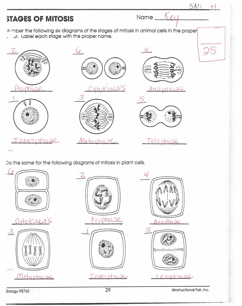
Animal Cell Simple Labeled Diagram - Mitosis Diagram Without Labels For ... The animal cell diagram is widely asked in class 10 and 12 examinations and is beneficial to understand the structure and functions of an animal. The diagram, like the one above, will include labels of the major parts of an animal cell including the cell membrane, nucleus, ribosomes, mitochondria, vesicles, and cytosol. mitosis_1_answers.pdf - SW Science 10 Unit 1 Mitosis... - Course Hero Label the following diagram of mitosis of an animal cell. 2. During which stage of a cell's cycle do the replicated chromosomes thicken and become visible? ______________________ 3. In animal cells, which structure is thought to produce the spindle fibers that help separate the sister chromatids during anaphase? ______________________ 4. Mitosis In Plant Cell Diagram Structure : Functions and Diagram Mitosis In Plant Cell Diagram. •Mitosis is a carefully controlled process that organizes and separates the chromosomes correctly. Karyokinesis followed by division of cytoplasm i.e. We all do not forget that the human body is quite elaborate and a method I discovered to are aware of it is by way of the way of human anatomy diagrams. Animal Cell Telophase Diagram : The Diagram Given Below Represents A ... Venn diagram comparing plant cells and animal file size: Animal cell diagram simple gcse. Some students may be able to identify some of the structures. Cytoplasm, ribosomes, rough endoplasmic reticulum; The red blood cells make up the blood, while the nerve cells make up the nervous system tissues. The correct diagram of animal cell is.
Animal Cell Mitosis Label Me! Printout - Enchanted Learning Metaphase - the phase of mitosis in which the chromosomes line up at the equator (the central plane) of the cell. Prophase - the phase of mitosis in which the duplicated chromosomes condense, the nuclear envelope dissolves, and centrioles divide and move to opposite ends of the cell. Mitosis (Definition, Diagram & Stages Of Mitosis) - BYJUS Mitosis Diagram showing the different stages of mitosis Mitosis is the phase of the cell cycle where the nucleus of a cell is divided into two nuclei with an equal amount of genetic material in both the daughter nuclei. It succeeds the G2 phase and is succeeded by cytoplasmic division after the separation of the nucleus. Label Plant And Animal Cells Teaching Resources | TpT Plant and Animal Cells Color and Label Parts. by. Rulers and Pan Balances. 41. $1.00. PDF. The set includes two versions of a plant cell to color and label and two versions of an animal cell to color and label. Students are ask to label the following organelles: nucleus cell wall cell membrane cytoplasm chloroplast mitochondrion endoplasmic ... Draw a well labelled diagram to show the anaphase stage of mitosis in a ... Draw a neat diagram of anaphase of mitosis of an animal cell and label the Polar region. 177236395. 3.3 k+. 9.4 k+. ... Draw a neat diagram of metaphase stage in an animal mitosis cell division and label the following parts : continuous spindle fibre,polar region,chromosome. chromosomal fibre. 643973380.
Cell Cycle Label | Biology worksheet, Cell cycle, Mitosis
03 Label the Cell Diagram | Quizlet Start studying 03 Label the Cell. Learn vocabulary, terms, and more with flashcards, games, and other study tools. ... 03 Cell Cycle and Mitosis. 16 terms. muskopf1 TEACHER. Sets with similar terms. 7.3 Structures of organelles ... 16 terms. schoolaccount40657. Anatomy : Cell functions. 14 terms. Kelly_Dng. cell diagram. 18 terms. lugo_janet ...

"Mitosis
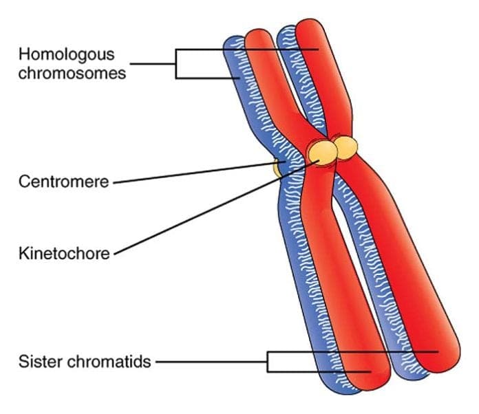
What Is A Homologous Chromosome? | Biology Explorer
33 Blank Animal Cell Diagram To Label Pdf - Labels Database 2020
i draw a diagram of the nucleus of a cell having chromosome number 6 as ...
Document
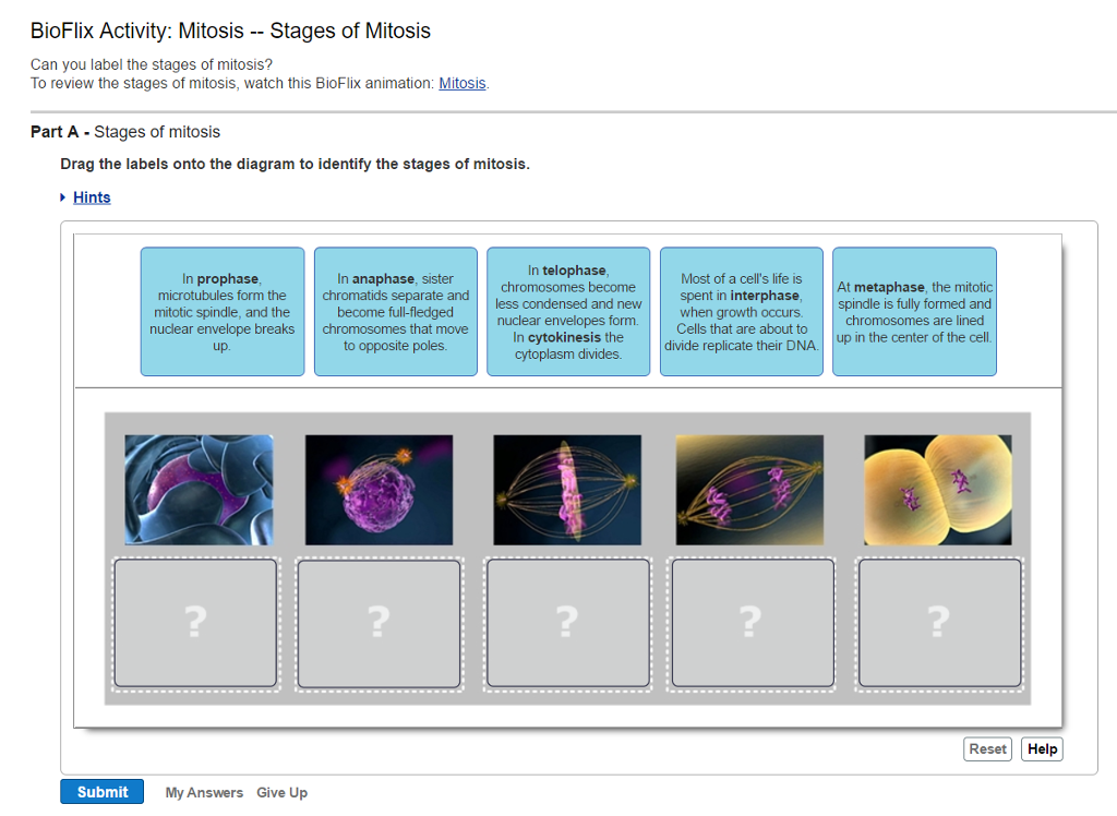
Solved: Can You Label The Stages Of Mitosis? To Review The... | Chegg.com

Cell Division Drawing at GetDrawings | Free download

Meiosis II - Stages and Significance of Meiosis-II Cell Division
Post a Comment for "43 label the following diagram of mitosis of an animal cell"