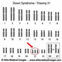
38 chromosome diagram unlabeled
Genetics Final (Multiple Choice) Flashcards | Quizlet A black dog is crossed with a brown one. In the first litter, all six puppies are black and one is brown. What is the best conclusion?, 3-4 5-6 1 2 Which pair(s) of numbers in the diagram identify(ies) homologous chromosomes?, 3-4 5-6 1 2 Which pair(s) of numbers in the diagram identify(ies) sister chromatids? and more. Bacteria in Microbiology - shapes, structure and diagram Bacterial spores. Bacterial endospores layers. Bacteria cells are the smallest living cells that are known; even though viruses are smaller than bacteria, viruses are not living cells. There are different types of bacteria with various sizes, shapes, and structures. The bacteria shapes, structure, and labeled diagrams are discussed below.
A Labeled Diagram of the Plant Cell and Functions of its Organelles The nucleus is known to be the 'control room' of the cell. It regulates various cell functions by controlling the protein synthesis of the plant cell. The nucleus contains DNA within the chromosomes. It is a membrane-bound structure that contains the cells hereditary information. Function: Controls expression and transcription of the gene ...

Chromosome diagram unlabeled
Diagram Lists - Stevenson High School You may wish consider printing the unlabeled diagrams, and labeling the significant structures yourself as you read or as diagrams are discussed in class. As an AP student, you are expected to be an active participant, so bring all diagrams to class for every unit. ... 12.4: Chromosome distribution and duplication during cell division 12.5: The ... hillis2e_ch09 - LaunchPad | Macmillan Learning for Instructors Each eukaryotic chromosome is composed of a double-stranded DNA molecule. It is possible to use a microscope to distinguish between a chromosome that has been labeled with radioactivity and one that is unlabeled. Plant cells were grown in the lab in the presence of radioactive thymidine for many generations. Single sister labeling in C. elegans chromosomes Chromosome axis ... Download scientific diagram | Single sister labeling in C. elegans chromosomes Chromosome axis (HTP-3) is labeled in red, EdU is in green and DAPI in blue. (A) A bivalent chromosome with two EdU...
Chromosome diagram unlabeled. DOC Mitosis: Labeled Diagram - West Branch High School The chromosomes line up separately on the spindle for metaphase of mitosis, but at the first division of meiosis the chromosomes pair in a process called synapsis. In synapsis, homologous chromosomes pair tightly along their length and then move to opposite poles so that only one of each pair of chromosomes ends up in each cell after the first ... Labeling Oligonucleotides and Nucleic Acids—Section 8.2 Unlabeled aminoallyl derivatives of dUTP and UTP, as well as unlabeled and labeled aminohexylacrylamide ... including chromosome and mRNA fluorescence in situ hybridization ... Figure 8.2.8 Schematic diagram of the labeling method provided in our ARES DNA Labeling Kits. The ARES DNA Labeling Kits use a two-step method to label DNA. PDF Meiosis Notes - sedelco.org Meiosis is a process in which the number of chromosomes per cell is cut in half through the separation of homologous chromosomes in a diploid cell. Meiosis usually involves two distinct divisions, called meiosis I and meiosis II. By the end of meiosis II, the diploid cell becomes four haploid cells. MeiosisLesson Overview Phases of Meiosis Animal Cell Telophase Diagram / Cell division...Prophase, Prometaphase ... Learners need to know the names of the phases and they need to be able to draw simple descriptive diagrams showing the chromosome changes. Under the microscope, an animal cell shows many different parts called organelles, that work together to keep the cell functional. ... Unlabeled animal cell diagram finally an unlabeled version of the ...
Human Heart Diagram Labeled - Science Trends Anatomy Of The Heart. The human heart usually weighs somewhere between 10 to 12 ounces in men and between 8 to 10 ounces in women, and in terms of size is roughly the size of the fist. The heart has four different chambers: the left and right ventricles and the left and right atriums. Of The Best Cell Cycle Diagram Unlabeled The graphic below shows an unlabeled animal cell. Image shows the stages of the cell cycle interphase prophase metaphase anaphase and telophase and asks students to name the phase and identify major structures such a centrioles and chromatids. Cells in human body. Diagram of Chromosome Structure - Online Biology Dictionary The name chromosome, meaning "colored body," is derived from the fact that in early studies of cellular structure the chromosomes could be easily stained with colored dyes and therefore showed up as colored bodies under the microscope. The diagram of chromosome structure above shows how DNA is organized in a eukaryotic cell. Candidate genes in genomic regions recurrently affected by focused ... B, C Minimally amplified regions (indicated in vertical lines) for amplifications of chromosome 5 with candidate genes. The filled circles on the left side indicate mucosal melanomas, with ...
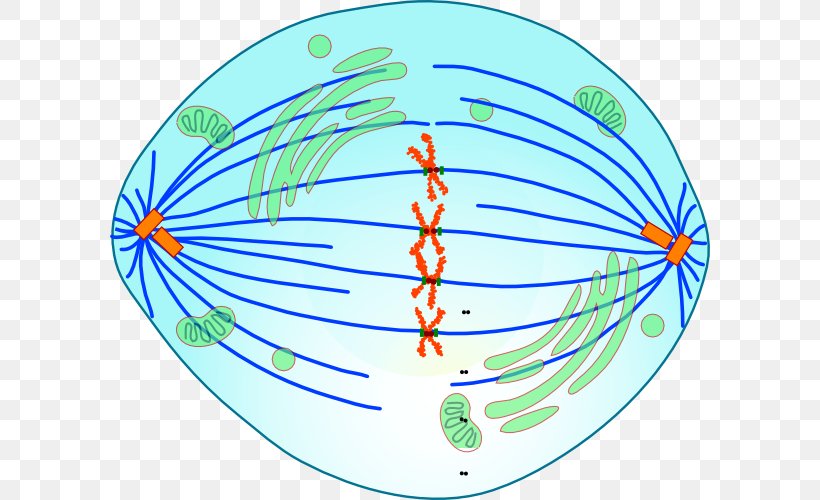
Solved Transcribed image text: Given the diagram below, assume that a G1 chromosome (e underwent one round of replication in thymidine and the metaphase chromosome (right) had both chromatids labeled. Which of the following replicative models conservative depressive seniservative could be eliminated by this observation? Mitosis | Biology I Laboratory Manual | | Course Hero In this exercise, we will consider prometaphase a component of prophase. Part 1. Mitosis Diagram Using Figures 1-4, diagram the phases of mitosis and in the space provided to the right, describe the events of each phase using a chromosome number of 6 (2n = 6). Figure 1. Prophase Figure 2. Metaphase Figure 3. Anaphase Figure 4. Telophase Draw And Label Diagrams Of Plant Cell And Animal Cell : Printable ... Cells form the basic building blocks for all living things. Centrioles help move chromosomes during cell division. A comparison of plant and animal cells using labelled diagrams and descriptive explanations. Label the plant cell diagram using the glossary of plant cell terms. A system of flattened membranes called cisternae (mainpoint: Chromosome Clipart Teaching Resources | Teachers Pay Teachers 2. $3.00. PDF. Google Apps™. These interactive diagrams are terrific for science teachers teaching their students about Chromosome and Chromosome Structure in a distance learning or blended learning environment. These click-and-learn diagrams are Google Slides files designed to be used in several possible ways.*.

What is a Chromosome? - B4FA
Unlabeled Animal Cell Diagram Free Simple Finally, an unlabeled version of the diagram is included at the bottom of the page, in color and black and white. Download Free HD Wallpapers [Mobile + Desktop] SEARCH. We all keep in mind that the human physique is very problematic and a method I discovered to comprehend it is by means of the way of human anatomy diagrams.

Alila Medical Media | Cell, Molecular Biology & Genetics Images
A Labelled Diagram Of Meiosis with Detailed Explanation Here, the chromosomes begin to condense. Prophase I is divided into five different stages: Leptotene Zygotene Pachytene Diplotene Diakinesis Metaphase I The homologous pairs of chromosomes are aligned on the equatorial plate. Anaphase I The homologous chromosomes are pulled on the opposite poles. The sister chromatids remain attached to each other.
genetic Archives - Medical Information Illustrated
The Structure of an Atom Explained With a Labeled Diagram Basic Diagram of an Atom. Most of an atom is just empty space and consists of a positively charged nucleus of protons and neutrons surrounded by a cloud of negatively charged electrons. The center of an atom is the nucleus and one or more electrons surrounding the nucleus. When one says an atom is electrically neutral, it means that the number ...

Biology Pictures: The Cardiac Cycle Picture
Structure of Chromosomes (With Diagram) | Cell Nucleus | Biology Each chromosome consists of one to four coiled threads called chromonema and also contains juxtaposed minute particles known as chromomeres (Fig. 1.15) which are rich in DNA. Most of the chromosomes possess usually two constrictions - primary (kinetochore) and secondary.

Ribosomes Images, Stock Photos & Vectors | Shutterstock
Structure of Bacterial Cell (With Diagram) - Biology Discussion It is 10-25 nm in thickness. It gives shape to the cell. Nucleus: The single circular double-stranded chromosome is the bacterial genome. Other structures include cytoplasmic membrane, mesosomes, ribosomes and cytoplasmic inclusions. Unlike eukaryotes cytoplasm does not contain ribosome, Golgi, cytoskeleton.
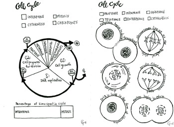
Cell Cycle and Mitosis coloring sheet by Scientifically Speaking is my ...
labeling a animal cell Printable Animal Cell Diagram - Labeled, Unlabeled, And Blank . unlabeled. ... mitosis cell cycle stages labeled labeling worksheet chromosome practice chromosomes biologycorner meiosis cells each humans resources structure worksheeto. How to make an animal cell : 10 steps. Animal cell worksheet.

Homologous Chromosomes Crossing Over 1 Clip Art at Clker.com - vector ...
Unlabeled Clip Art - Royalty Free - GoGraph Download high quality Unlabeled clip art from our collection of 66,000,000 clip art graphics. 800-810-1617 gograph@gograph.com ... Medical Labeled Diagram Closeup With Muscle, Transverse Carpal Ligament, Median Nerve, Tendon Sheath, Flextor Tendons And Bones. ... Educational And Medical Scheme With Cell, Chromosome And Dna. Labeled Anatomical ...

Alila Medical Media | Cell, Molecular Biology & Genetics Images
Plant Cell Unlabeled Teaching Resources | Teachers Pay Teachers This resource is an assorted pack of activities to help students learn the structure and function of plant and animal cell organelles. A variety of diagrams both labeled and unlabeled are included as well as interactive notebook activities and foldables to reinforce the structure and function of plant and animal cell organelles.
Metaphase Mitosis Anaphase Meiosis Cell, PNG, 600x500px, Metaphase ...
Learn the parts of a cell with diagrams and cell quizzes - Kenhub For this exercise we'll start with an image of a cell diagram ready labeled. Study this and make sure that you're clear about which structure is found where. Cell diagram unlabeled It's time to label the cell yourself! As you fill in the cell structure worksheet, remember the functions of each part of the cell that you learned in the video.
Post a Comment for "38 chromosome diagram unlabeled"