43 dissected cow eye labeled
PDF Cow Eye Dissection Guide - Central Bucks School District DISSECTION OF THE COW EYE Please make sure to wear gloves and safety glasses when you are dissecting, and make sure to clean up thoroughly after the lab. Also, the cow eyes can be rather slippery, so use caution when handling and cutting them. You will need a scalpel and forceps. 1. First, identify the most external structures of the eye. Texas Eye in Garland, TX with Reviews - YP.com Find 1037 listings related to Texas Eye in Garland on YP.com. See reviews, photos, directions, phone numbers and more for Texas Eye locations in Garland, TX.
EOF

Dissected cow eye labeled
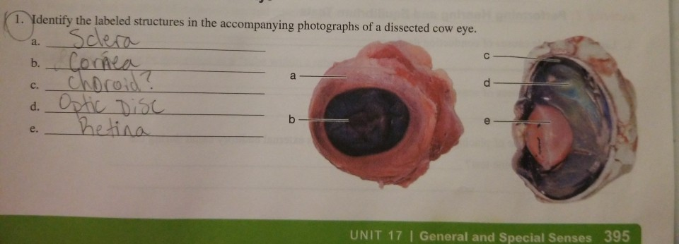
Times & The Sunday Times Jul 22, 2022 · News and opinion from The Times & The Sunday Times › 40252505 › Bancrofts_Theory_andBancroft's Theory and Practice of Histological Techniques Supersensitive In Situ Hybridization by N Tyramide Signal Amplification and Nanogold@ Silver Staining: The Contribution of Autometallography and Catalyzed Reporter Deposition to the Rejuvenation of In Situ Hybridization Solved 1. Identify the labeled structures in the | Chegg.com Anatomy and Physiology questions and answers. 1. Identify the labeled structures in the accompanying photographs of a dissected cow eye. a. Sclera b. Cornea c. choroid? d. Optic Disc - Retina UNIT 17 | General and Special Senses 395. Question: 1. Identify the labeled structures in the accompanying photographs of a dissected cow eye. a. Sclera b.
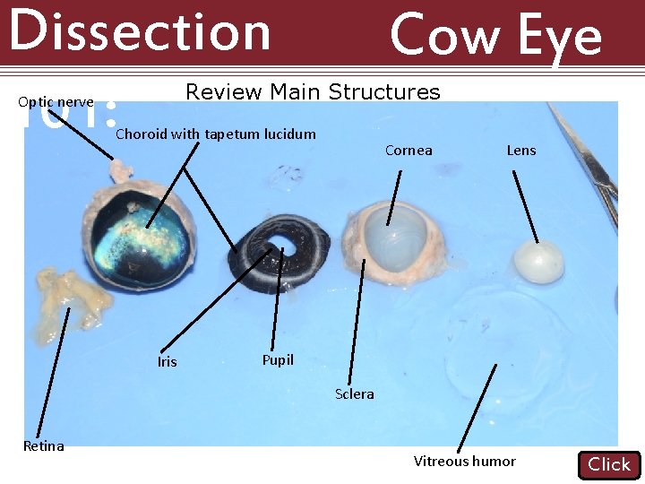
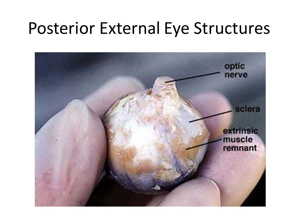
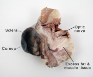
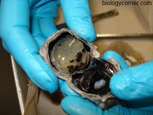
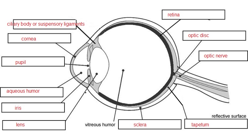
Dissected cow eye labeled. › news › giant_hogweed_not_widelyGiant hogweed: Not widely spread in Michigan - Landscaping Aug 04, 2015 · A member of the carrot and Queen Anne’s lace family, giant hogweed exhibits many of the same characteristics of other members of this group such as cow parsnip and angelica, including having sap that can cause severe dermatitis. The plant bears chunky, greenish-purple stems topped by platter-sized, dissected foliage. Cow Eye Dissection & Anatomy Project | HST Learning Center Cow Eye Dissection: Internal Anatomy 1. Place the cow's eye on a dissecting tray. The eye most likely has a thick covering of fat and muscle tissue. Carefully cut away the fat and the muscle. As you get closer to the actual eyeball, you may notice muscles that are attached directly to the sclera and along the optic nerve. Cow's Eye Dissection - Eye diagram - Exploratorium A cow's iris is brown. many colors, including brown, blue, green, and gray. A clear fluid that helps the cornea keep its rounded shape. The pupil is the dark circle in the center of your iris. It's a hole that Your pupil is round. is oval. A tough, Light bends as it passes through the cornea. PDF Cow Eye Dissection Lab - Home Science Tools This cow eye dissection kit comes with everything you need to conduct a lab examination. Safety Guidelines • Work in a place separate from eating and food preparation areas. • Use disposable latex gloves or nitrile gloves during the dissection and cleanup. • Use only dissection tools provided.
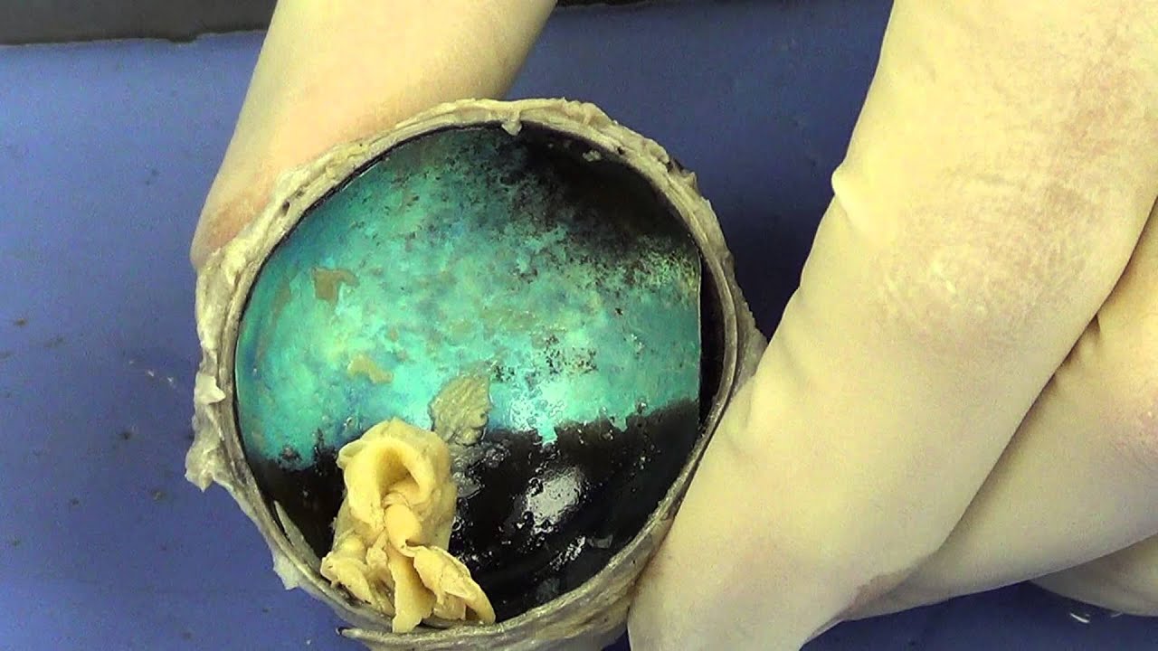
en.wikipedia.org › wiki › Wikipedia:Unusual_articlesWikipedia:Unusual articles - Wikipedia A Jersey cow that produced record amounts of butter and got a sizable neighborhood named for her. Lin Wang: A Taiwanese elephant made famous for his participation in the Second Sino-Japanese War. Lion of Gripsholm Castle: What happens when you tell a taxidermist who doesn't know what a lion is to stuff and mount a lion. Lonesome George PDF COW'S EYE dissection - Exploratorium COW'S EYE dissection page 6 Now take a look at the rest of the eye. If the vitreous humor is still in the eyeball, empty it out. On the inside of the back half of the eyeball, you can see some blood vessels that are part of a thin fleshy film. That film is the retina. Before you cut the eye open, the vitreous humor › book › showA Single Man by Christopher Isherwood - Goodreads Mar 20, 2001 · “But now isn’t simply now. Now is also a cold reminder: one whole day later than yesterday, one year later than last year. Every now is labeled with its date, rendering all past nows obsolete, until — later of sooner — perhaps — no, not perhaps — quite certainly: it will come.” — 164 likes Solved Activity 3: Dissecting the Mammalian Eye 1. Identify - Chegg Science. Anatomy and Physiology. Anatomy and Physiology questions and answers. Activity 3: Dissecting the Mammalian Eye 1. Identify the labeled structures in the accompanying photographs of a dissected cow eye b. C. d. e. Activitv 4: Performine Vicual Tectc.
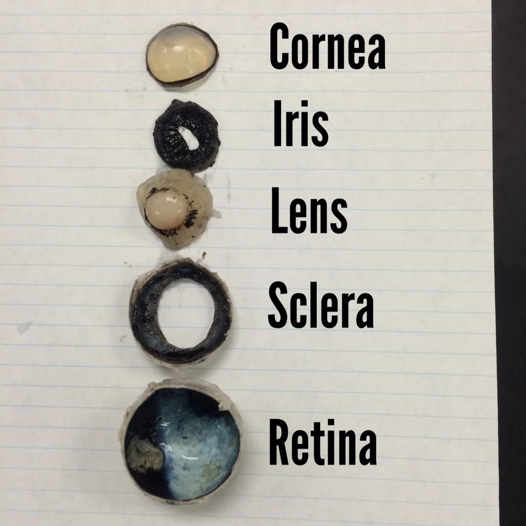
Cow Eye Dissection & Parts of the Eye Diagram | Quizlet cornea Clear, outer layer of the front of the eye. sclera White, outermost layer of the eye. Helps maintain shape and gives attachment to muscles. photoreceptors The cells in the retina that respond to light (rods and cones) rods Photoreceptor cells in the eye that detect black, white, and gray cones Photoreceptor cells in the eye that detect color Cow Eye Dissection - The Biology Corner 1. Examine the outside of the eye. You should be able to find the sclera, or the whites of the eye. This tough, outer covering of the eyeball has fat and muscle attached to it 2. Locate the covering over the front of the eye, the cornea. When the cow was alive, the cornea was clear. In your cow's eye, the cornea may be cloudy or blue in color. 2. Dallas Eye Associates - Eye Care Clinic in Dallas, TX Dallas Eye Associates is a Optometrist Center in Dallas, Texas. It is situated at 6500 Greenville Ave, Suite 150, Dallas and its contact number is 214-692-1901. The authorized person of Dallas Eye Associates is Dr. James Daniel Penick who is Provider/proprietor of the clinic and their contact number is 214-692-1901. Primary license number for Dallas Eye Associates is 0187OT (Optometrist) in Texas. PDF Cow Eye Dissection: Examining Structure and Function The eyes of cows are structurally and functionally similar to the eyes of humans. During this activity, you will dissect a cow eye. You will observe several important features of the eye and develop your understanding of how each part functions to make vision possible. Materials
Detailed Cow Eye Dissection: Part II (Jr. High, High School and College Review)
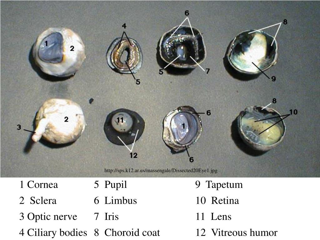
Cow Eye Dissection Guide - Google Slides Cow Eye. Use the point of a scissors or a scalpel to make an incision through the layers of the eye capsule (similar to figure 1); there are three layers from the exterior: sclera, whitish/grey, continuous with the transparent cornea, choroid, thin dark black layer and the retina, thin greyish/pink layer. Use a scissors to dissect the entire ...

Cow Eye Dissection | Perkins eLearning
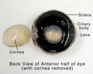
Cow eye - dissection and label Cow eye-shown with labeled Sclera The sclera (from the Greek skleros, meaning hard [1]), also known as the white of the eye, is the opaque, fibrous, protective, outer layer of the eye containing collagen and elastic fiber. 4. Cow eye-shown with lens dissected.
Solved 1. Identify the labeled structures in the | Chegg.com
Gulf Tape Label Co in Dallas, TX with Reviews - YP.com Find 2 listings related to Gulf Tape Label Co in Dallas on YP.com. See reviews, photos, directions, phone numbers and more for Gulf Tape Label Co locations in Dallas, TX.

COW EYE DISSECTION - NANDINI SONI
Lab: Cow Eye Dissection Flashcards | Quizlet the white outer layer of the eyeball. At the front of the eye it is continuous with the cornea aqueous humor transparent, watery fluid similar to plasma, but containing low protein concentrations. It is secreted from the ciliary epithelium, a structure supporting the lens vitreous humor
NASCOGuard®, Cow Organ - Eye, Preserved LS01628|Nasco
recorder.butlercountyohio.org › search_records › subdivisionWelcome to Butler County Recorders Office Copy and paste this code into your website. Your Link Name

Live Science Cow Eye Dissection – Otosection
Asd Label Inc. - Company Profile Asd Label Inc. filed as a Domestic For-Profit Corporation in the State of Texas on Monday, August 5, 2019 and is approximately three years old, according to public records filed with Texas Secretary of State. Sponsored Learn More D&B Reports Available for Asd Label Inc. Network Visualizer ...
PPT - COW EYE DISSECTION PowerPoint Presentation, free ...
learning-center.homesciencetools.com › articleSheep Brain Dissection Project Guide | HST Learning Center Sheep brains, like other sheep organs, are much smaller than human brains but have similar features. They can be a valuable addition to your study of anatomy. See for yourself what the cerebrum, cerebellum, spinal cord, gray and white matter, and other parts of the brain look like with this sheep brain dissection guide!

Cow Eye Dissection Guide - Google Slides
Cow Eye Dissection Kit for Kids Animal Anatomy Labs | HST Cow Eye Dissection Kit Cow Eye Dissection Kit $9.95 This Cow Eye Dissection Kit gives an inside view of how the eye works. It comes with everything you need for this activity, including a preserved cow eye specimen, a step-by-step dissection guide & essential dissection tools. quantity Ages 11+ In Stock & Ready to Ship Need it fast?
Cow Eye Dissection
Solved 1. Identify the labeled structures in the | Chegg.com Anatomy and Physiology questions and answers. 1. Identify the labeled structures in the accompanying photographs of a dissected cow eye. a. Sclera b. Cornea c. choroid? d. Optic Disc - Retina UNIT 17 | General and Special Senses 395. Question: 1. Identify the labeled structures in the accompanying photographs of a dissected cow eye. a. Sclera b.

Cow Eye Dissection Kit | Anatomy Warehouse
› 40252505 › Bancrofts_Theory_andBancroft's Theory and Practice of Histological Techniques Supersensitive In Situ Hybridization by N Tyramide Signal Amplification and Nanogold@ Silver Staining: The Contribution of Autometallography and Catalyzed Reporter Deposition to the Rejuvenation of In Situ Hybridization

Cow Eye Dissection & Labeling
Times & The Sunday Times Jul 22, 2022 · News and opinion from The Times & The Sunday Times
Student Mr Zoras on Twitter: "Great cow eye dissection today ...

Carolina® Eye Dissection Mat

Cow Eye Dissection Diagram | Quizlet

parts of the eye | Medical anatomy, Eye anatomy, Anatomy and ...
Lab - Eye Dissection: MAH-Summer 2019-Anatomy and Physiology I

Introduction into the Bovine eye and diagnostic and
SCB209 - Lab3 - Natural Sciences Open Educational Resources
Cow Eye Dissection & Anatomy Project | HST Learning Center
Cow Eye Dissection Teaching Resources | Teachers Pay Teachers
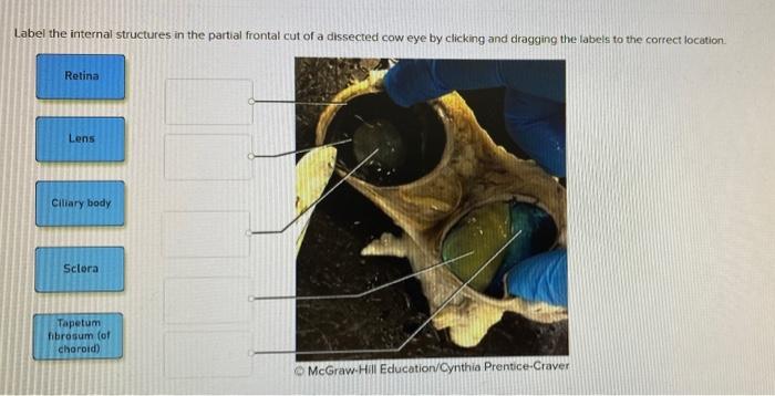
Solved Label the internal structures in the partial frontal ...

Cow Eye Dissection | Carolina.com
NEUR 320: Art and Vision
Dissection 101 Reasons to Use the Dissection Video
Sheep Eye Dissection. - ppt video online download

Cow Eye Dissection | Carolina.com

Cow Eye

Eye Dissection
Cow Eye Dissection & Anatomy Project | HST Learning Center
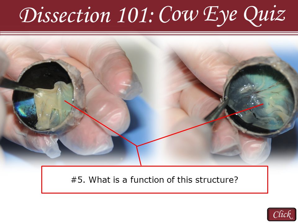
Cow Eye Quiz Dissection 101: Click - ppt video online download
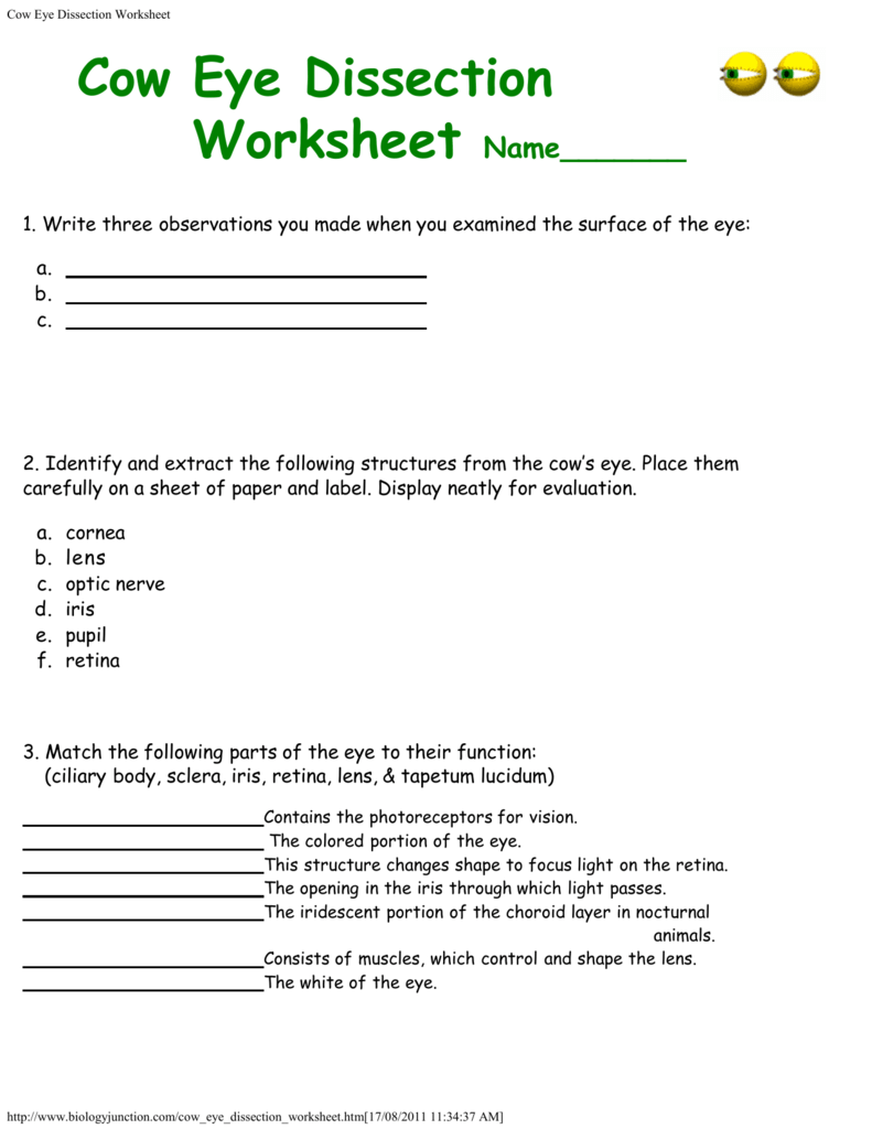
Cow Eye Dissection Worksheet

Eye Dissection Dissection Biology Classroom Anatomy – Otosection
Cow Eye Dissection

Cow Eye Dissection 2 Diagram | Quizlet

Young Scientist's Eye Dissection Kit: Science Lab Biology ...
Cow Eye Dissection

Dissection Cattle Anatomy Human eye, luminescent blue glow ...

Cow's Eye Dissection - Eye diagram

Cow Eye Dissection | Carolina.com

Cow's Eye Dissection | Exploratorium Video

Sheep Eye Disection | Mohtadi Alkhaliq

Cow Eye Dissection Post Lab - Aidan - Eye Dissection Post Lab ...
Cow Eye Dissection: Examining Structure and Function
Post a Comment for "43 dissected cow eye labeled"