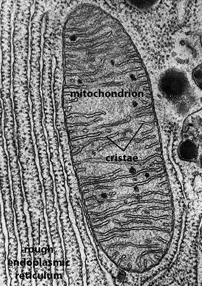
38 label the transmission electron micrograph of the mitochondrion.
(Get Answer) - Which structure is highlighted? O goblet ... - Transtutors Label the transmission electron micrograph of the cell. 0 Nucleus rences Mitochondrion Heterochromatin Peroxisome Vesicle ULAR bumit Click and drag each label into the correct category to indicate whether it pertains to the cytoplasm or the plasma... Identifying long-range synaptic inputs using genetically encoded labels ... Top, labeled and unlabeled mitochondria have significantly different pixel values (p-value = 4.76 × 10-95, n = 148, unlabeled, green; n = 255, unlabeled, gray). ( k) Mitochondria mean pixel value...
Multiclass U-Net Segmentation of Brain Electron Microscopy ... - Springer Open EM data as a whole are represented by only a few labeled dataset, both due to the laboriousness of preparing samples for an electron microscope, and due to the lack of specialists for manual labeling. We found four open EM datasets the earliest and most popular of which are labeled only for one class (mitochondria or membranes).

Label the transmission electron micrograph of the mitochondrion.
EOF Course: s4: Biology , Topic: UNIT 3: MICROSCOPY Observe, draw and label the visible parts under a light microscope. Avail these materials before you start: Petri-dishes, plate covers, pencil, transparent tape, microscope, agar powder, and permanent slide of bacteria, amoeba, and paramecium, Bunsen burner or any other source of heat. Procedure Label This Transmission Electron Micrograph / Microscopy Innovations ... Label the transmission electron micrograph of the nucleus. 0 nucleus rences mitochondrion heterochromatin peroxisome vesicle ular bumit . Scanning Transmission Electron Micrograph Stem Vesicles Were Download Scientific Diagram from Labeling nuclear proteins with electron dense probes in living cells. 0 nucleus rences ...
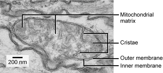
Label the transmission electron micrograph of the mitochondrion.. Transmission electron microscopy study of the cell-sensor interface Typical scanning electron microscope images of platinum/coal-sputtered epoxy imprints of the FET surface and one single FET gate after the mechanical removal of the chip. One can clearly identify the well-preserved imprints of the contact lanes and gates of a FET chip. Scale bars, ( a) 500 μm and ( b) 10 μm. Laboratory Manual for Non-Majors Biology - Google Books Result James W. Perry, David Morton, Joy B. Perry · 2012 · ScienceIdentify and label the outer membrane separating the organelle from the cytoplasm. ... Figure 10-6 Transmission electron micrograph of a mitochondrion (18 ... (Get Answer) - Label this transmission electron micrograph of relaxed ... Label this transmission electron micrograph of relaxed sarcomeres by clicking and dragging the... Label this transmission electron micrograph of relaxed sarcomeres by clicking and dragging the labels to the correct location Sarcamere 1 band (light) Z disc Mline Aband (dark) H zone. Oxidative stress-mediated mitochondrial fission promotes hepatic ... F, G Representative transmission electron microscopy images and quantification of the mitochondrial dynamics in livers from olive oil- and CCl 4 -treated mice. Scale bar, 2 μm. Mitochondria are...
Condensed Mitochondria Assemble Into the Acrosomal Matrix During ... Transmission Electron Microscopy Testes of mice were de-capsulated and fixed with 2.5% glutaraldehyde and 2% paraformaldehyde in 0.1M sodium cacodylate buffer (pH 7.2) for 2 h. After treatment with 1% osmium tetroxide for 1 h, the testes were stained with 1% uranyl acetate aqueous solution overnight at 4°C. In-resin CLEM of Epon-embedded cells using proximity labeling When mitochondria-localized miniTurbo was expressed in the cells, mitochondria-like fluorescent signals were detected in the sections, and ultrastructures of mitochondria were observed as fluorescence-positive structures in the same sections by scanning electron microscopy. Proximity labeling using miniTurbo led to more stable and brighter ... Plant Cell Nucleus Electron Micrograph : Cell And Organelles Dr Jastrow ... This electron micrograph shows a mitochondrion as viewed with a transmission electron microscope. The nucleus (plural = nuclei) figure 7.14 at left a transmission electron micrograph and at right a labeled diagram of a. An electron micrograph of a cell nucleus showing a densely staining nucleolus. Transmission Electron Microscope (TEM)- Definition, Principle, Images The working principle of the Transmission Electron Microscope (TEM) is similar to the light microscope. The major difference is that light microscopes use light rays to focus and produce an image while the TEM uses a beam of electrons to focus on the specimen, to produce an image. Electrons have a shorter wavelength in comparison to light which ...
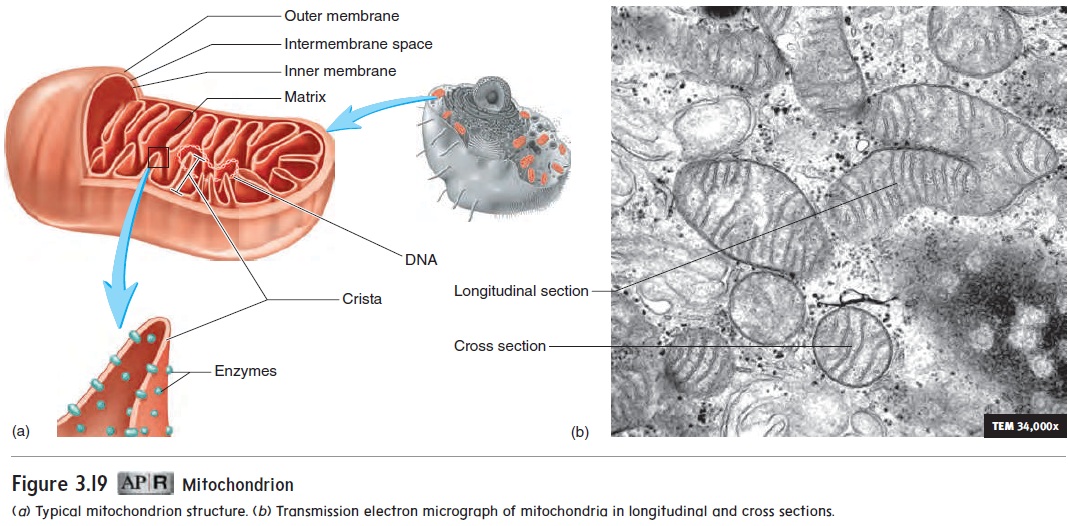
Plant Cell Electron Micrograph Labelled : Plant Bodies Cells / In this ... Below is a collection of electron micrographs with labelled subcellular structures that you should be able to identify. Figure 7.14 at left a transmission electron micrograph and at right a labeled diagram of a mitochondrion. Atp synthesis takes place on the inner membrane. Plant leaf tissue but not root. Label This Transmission Electron Micrograph : TEM of chloroplast from ... Label this transmission electron micrograph of relaxed sarcomeres by clicking and dragging the labels to the correct location . Transmission electron microscopy (tem) is one of the oldest technologies and still. Molecular labeling for correlative microscopy: Fluorescence microscopy in combination with tem and an ion beam analysis (iba, which ... Mitochondrion - Wikipedia A mitochondrion ( / ˌmaɪtəˈkɒndriən /; [1] pl. mitochondria) is a double- membrane -bound organelle found in most eukaryotic organisms. Mitochondria use aerobic respiration to generate most of the cell 's supply of adenosine triphosphate (ATP), which is subsequently used throughout the cell as a source of chemical energy. [2] Label This Transmission Electron Micrograph / Microscopy Innovations ... Label the transmission electron micrograph of the nucleus. 0 nucleus rences mitochondrion heterochromatin peroxisome vesicle ular bumit . Scanning Transmission Electron Micrograph Stem Vesicles Were Download Scientific Diagram from Labeling nuclear proteins with electron dense probes in living cells. 0 nucleus rences ...
3.3 Eukaryotic Cells – Concepts of Biology – 1st Canadian Edition
Course: s4: Biology , Topic: UNIT 3: MICROSCOPY Observe, draw and label the visible parts under a light microscope. Avail these materials before you start: Petri-dishes, plate covers, pencil, transparent tape, microscope, agar powder, and permanent slide of bacteria, amoeba, and paramecium, Bunsen burner or any other source of heat. Procedure

Mitochondrial Protrusions in Neuronal Cells - ScienceDirect
EOF

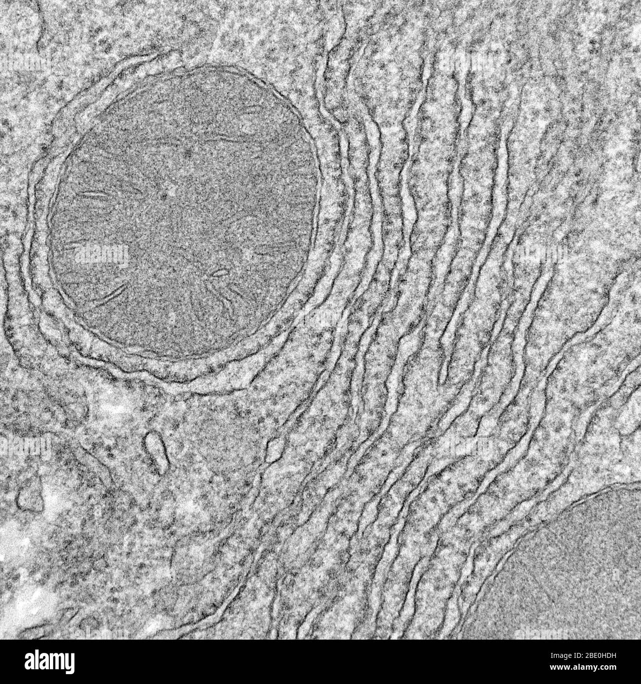
TEM of mitochondrian in cell - Stock Image - G465/0090 ...
Transmission Electron Microscopy of Biological Samples ...

Electron Microscopic Analysis of a Spherical Mitochondrial ...
Transmission Electron Micrograph (TEM) showing mitochondria ...

Mitochondrial morphology, topology, and membrane interactions ...
Ultrastructure of cells 1.2
BIOL 230 Lecture Guide - Electron Micrograph of Mitochondria
Toxins | Free Full-Text | The Phospholipase Activity of ...

Subpopulation-specific differences in skeletal muscle ...
Floral micromorphology, histochemistry, ultrastructure and ...
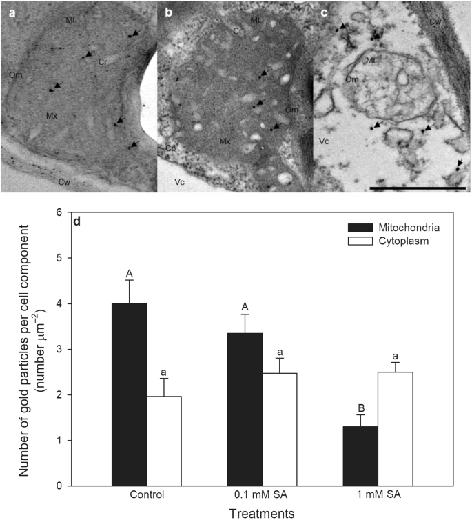
Salicylic acid-induced ROS production by mitochondrial ...
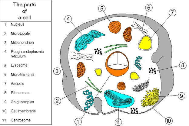
Organelles - Cell Structures and their Functions
Electron Micrographs

Pathways shaping the mitochondrial inner membrane | Open Biology
The mitochondrial compartment

The Cell. - ppt download

The morphology of mitochondria. (a) Thin-section electron ...
Electron Micrographs
A-level Biology Bridging Course - Week 1
Cambridge International Examinations Cambridge International ...
Frontiers | Mitochondrial Morphology and Mitophagy in Heart ...
9700 QR Dynamic Papers Biology al Cambridge
A Guide for Using Transmission Electron Microscopy for ...

Cell Micrographs | BioNinja
Biology | Free Full-Text | Pomacea canaliculata Ampullar ...
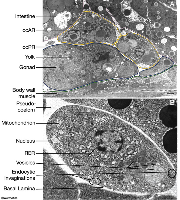
Hermaphrodite Coelomocyte System

Mitochondria-Associated Membranes: Composition, Molecular ...
Transmission Electron Micrograph (TEM) showing mitochondria ...

Structure & Function of Mitochondria (12.2.1) | CIE A Level ...
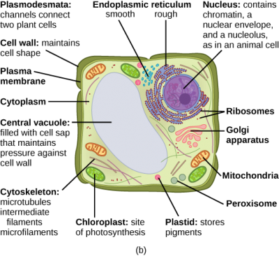
3.3 Eukaryotic Cells – Concepts of Biology – 1st Canadian Edition

Pin page
Topic 1.2 Ultra-Structure of Cells - AMAZING WORLD OF SCIENCE ...
Reading: Mitochondria | Biology (Early Release) | | Course Hero

Lewy pathology in Parkinson's disease consists of a crowded ...

2.3.3 Identify structures from electron micrographs of liver ...

Differential Alterations of the Mitochondrial Morphology and ...
Post a Comment for "38 label the transmission electron micrograph of the mitochondrion."