41 nucleus picture with labels
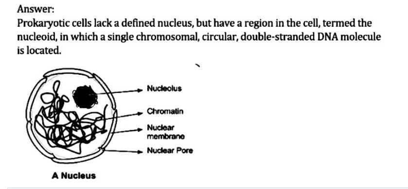
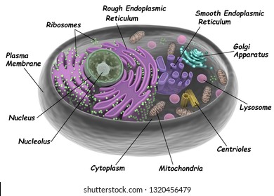
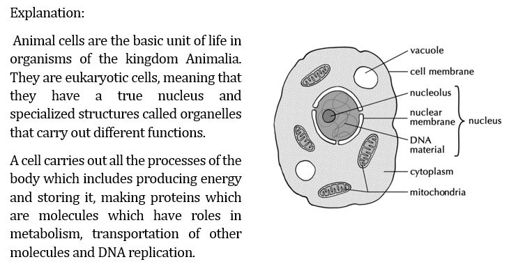
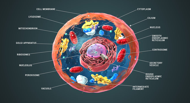
How to Draw an Animal Cell: 11 Steps (with Pictures) - wikiHow 1. Draw a simple circle or oval for the cell membrane. The cell membrane of an animal cell is not a perfect circle. You can make the circle misshapen or oblong. The important part is that it does not have any sharp edges. [1] Also know that the membrane is not a rigid cell wall like in plant cells. Nucleus- Definition, Structure, Parts, Functions, Diagram The cell nucleus is a membrane-bound structure that contains the cell's hereditary information and controls the cell's growth and reproduction. It is the command center of a eukaryotic cell and is commonly the most prominent organelle in a cell accounting for about 10 percent of the cell's volume. In general, a eukaryotic cell has only ...
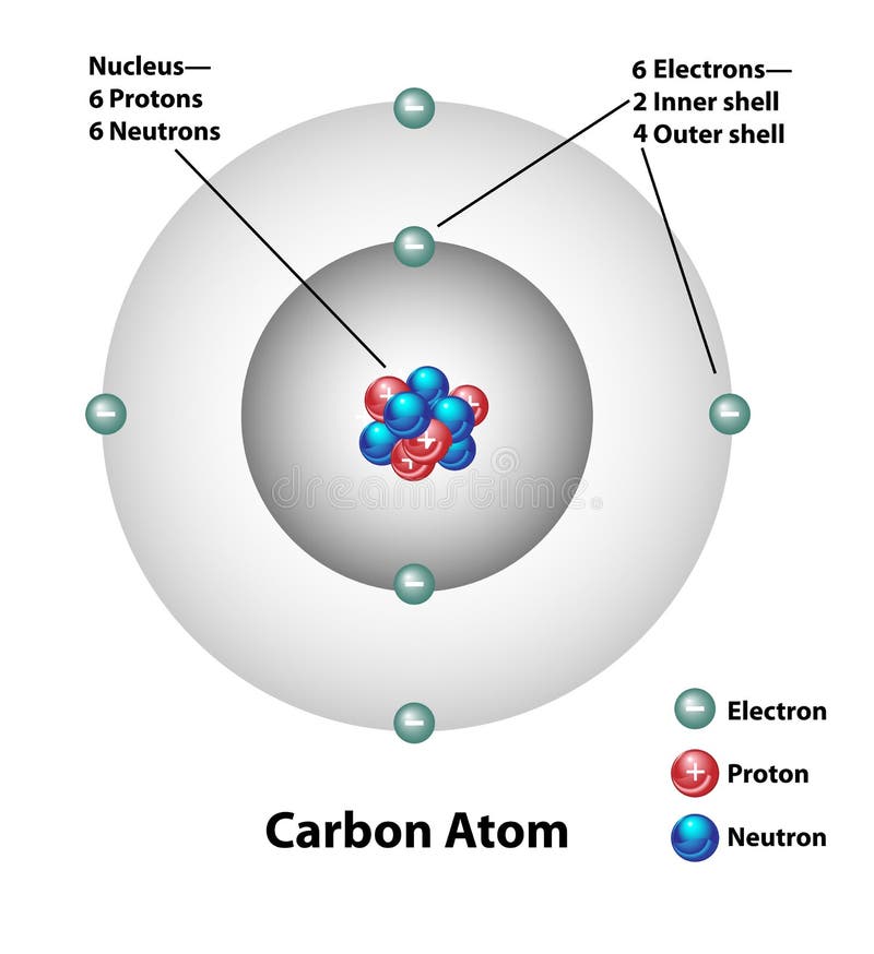
Basic Parts of the Atom - Protons, Neutrons, Electrons, Nucleus The Beautiful pictures of the atom in this video come from Jefferson Lab @ . They are here on youtube too @ ...
Nucleus picture with labels
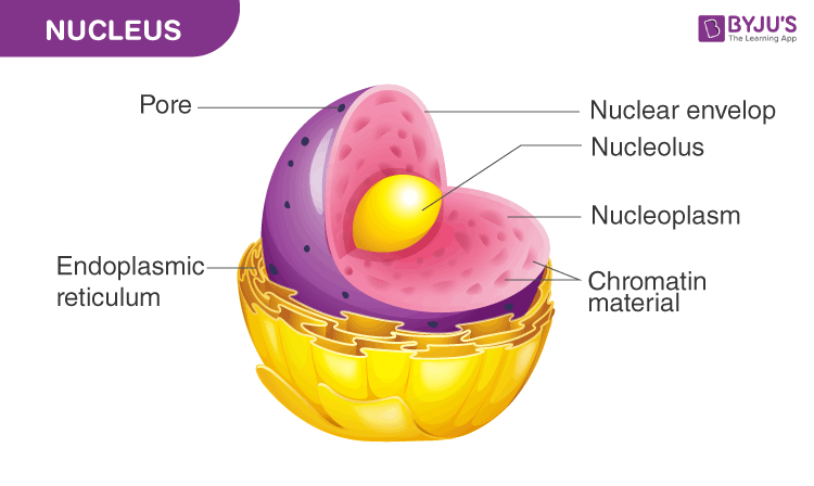
What is a Nucleus?- Structure and Function of Nucleus - Byju's The nucleus is a double-membraned organelle that contains the genetic material and other instructions required for cellular processes. It is exclusively found in eukaryotic cells and is also one of the largest organelles. Outline the structure of the Nucleus. A double-membraned organelle known as the nuclear membrane/envelope engirdles the nucleus. How to draw & label the nucleus - Pinterest The space between the inner and outer layers is called Pericardial space, it is filled with pericardial fluid. Pericardium and pericardial fluid protect the heart from physical shocks. Now lets start drawing the diagram. 1.Draw a oblique line from Right to Left 2.Draw a cone shape as shown in… J jessica Heart anatomy drawing Anatomy Drawing How to draw & label the nucleus - YouTube A beautiful drawing of the nucleus. And it will teach you draw Nucleus very easily. Watch the video and please be kind enough to thumbs up my videos. And I w...
Nucleus picture with labels. Cell Organelles and Function with Labels Flashcards | Quizlet Cell Organelles and Function with Labels. STUDY. Flashcards. Learn. Write. Spell. Test. PLAY. Match. Gravity. Created by. mollymorrison. Terms in this set (21) Cell. basic unit of all living things ... synthesized and partially assembled, located in the nucleus. Chromosome. a structure in the nucleus that contains hereditary material. Vacuole ... Animal Cell - Free printable to label + Color -kidCourses.com Can you label and color these important parts of the animal cell?. NUCLEUS control center for cell (cell growth, cell metabolism, cell reproduction). NUCLEOLUS synthesizes rRNA. RIBOSOMES the site of protein building, this is where translation takes place (mRNA in language of nucleic acids is translated into the language of amino acids). RER (Rough Endoplasmic Reticulum) synthesizes proteins ... Answered: Label cell wall, chromatin, and nucleus | bartleby Q: Label the nucleus. First picture is at 100x, second picture is at 400x. Q: Label cell membrane, nuclear membrane, chromatin, and use a bracker for the nucleus. Cheek cell is composed of epithelial cells and is isolated from the inner wall of the mouth. A cheek… Q: The period of the cell cycle when DNA replicates is _________________. a. Labeled Plant Cell With Diagrams | Science Trends Labeled Plant Cell With Diagrams Daniel Nelson 18, April 2019 | Last Updated: 3, March 2020 Plant cells contain many organelles such as ribosomes, the nucleus, the plasma membrane, the cell wall, mitochondria, and chloroplasts. In addition, plant cells differ from animal cells in a number of key ways.
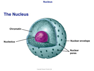
Animal Cells: Labelled Diagram, Definitions, and Structure Nucleus The nucleus is a highly specialized organelle that lives in its own double membrane with the nucleolus. The nucleus stores the cell's DNA and holds all the information the cell needs to do its job. Chromatin is a web-like substance that holds the nucleus's DNA. Nucleus Diagram, Definition, Structure and Function The nucleus is the command centre of a cell. This is because it contains the genetic material of the cell. Therefore, it consists of a number of structural elements which facilitate its functions. The nucleus of a cell has a spherical shape. A nucleus diagram is very useful for studying its structure. Its structure consists of the following ... Answered: Label the nucleus. First picture is at… | bartleby Label the nucleus. First picture is at 100x, second picture is at 400x. Question. thumb_up 100% Cell Illustrations - Cell nucleus (labels) - histology illustration FILE 32/55 Cell nucleus (labels) - histology illustration Diagram of an cell nucleus: 1. Nuclear envelope 1.a. Outer membrane 1.b. Inner membrane 2. Nucleolus 3. Nuceleoplasm 4. Chromatin 4.a. Heterochromatin 4.b. Euchromatin 5. Ribosomes 6. Nuclear pore Histlogy illustration courtesy of Mariana Ruiz.
Solved Art-labeling Activity: Neuron Structure 6 of 36 - Chegg Anatomy and Physiology questions and answers. Art-labeling Activity: Neuron Structure 6 of 36 Review Part A Drag the labels to the appropriate location in the figure. Axon Nucleus Synaptic terminals Microfibrils and microtubules CO on Coll body II. Mitochondrion Dendrites Nucleolus Submit Request Answer Solv cheo. Human Cell Diagram, Parts, Pictures, Structure and Functions The nucleus is the master control of the cell. It contains genes, collections of DNA, which determines every aspect of human anatomy and physiology. The DNA which is arranged into chromosomes also contains the blueprint specific for each type of cell which allows for replication of the cell. Within the nucleus is an area known as the nucleolus. Chapter 3 Flashcards | Quizlet Label the parts of the nucleus. Osmosis is the movement of. water molecules from a high concentration to a low concentration through a selectively permeable membrane. ... Drag the descriptions of plasma membrane extensions to the appropriate picture of cilia and/or flagella.Labels can be used more than once. The Structure of an Atom Explained With a Labeled Diagram The atomic model in the diagram below shows protons and neutrons concentrated at the atomic nucleus and electrons in the orbits surrounding it. Protons are positively charged, electrons are negatively charged, while the neutrons carry no charge. J.J. Thomson Plum Pudding Model
Chapter Seven- The Cell
The Structure of an Atom: Model, Diagram, Examples - Embibe The nucleus is positively charged since the proton is positively charged and the neutron is neutral. 4. The electrons are negatively charged. 5. Atoms are neutral as a whole. Q.3. How do you know the structure of an atom? Ans: The structure of an atom can be known by the atomic number and mass number. The atomic number is equal to the number of ...
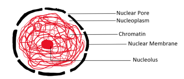
Draw a Nucleus and label any 4 parts. Name the type of ...
Nucleolus Photos and Premium High Res Pictures - Getty Images nucleus ribosome cell cell membrane cytoplasm endoplasmic reticulum lysosome nucleous nuclear pore vacuole 118 Nucleolus Premium High Res Photos Browse 118 nucleolus stock photos and images available, or search for nucleus or ribosome to find more great stock photos and pictures. Related searches: nucleus ribosome cell cell membrane cytoplasm of 2
Simple Atom Symbol, Molecule Concept, Structure of the ...
Screenbound Pictures / Odeon Entertainment - Page 88 - Cult Labs Location: Yorkshire. Quote: Originally Posted by peter alex. the libertine and The Garden of Torment was released by Nucleus Films not Screenbound pictures/Odeon entertainment. Screenbound also stock titles from various other labels, such as Severin and Nucleus, so I think I can see why you were confused. Share.

Binary Fission Diagram with Labels Stock Vector ...
1. What cell types are illustrated in above picture? 2. Label the... 2. to demarcate the regions you can use those dotted lines. On a conceptual level a protein syntesized by mRNA is always in the cytoplasm and TFs (transcription factors) will be inside nucleus so use them for your reference as well. 3. Protein L in Liver cells Protein H in Hair cells
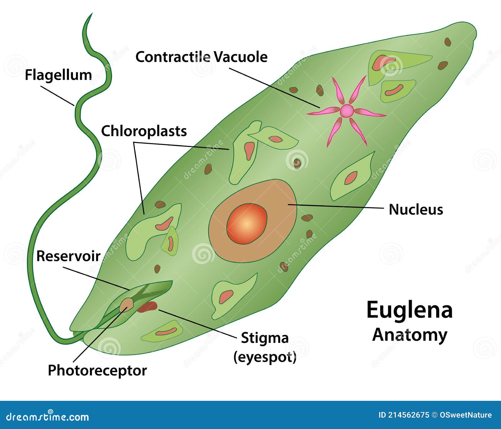
Euglena Protozoan Microscopic Cell Structures Stock Vector ...
Label An Atom Teaching Resources | Teachers Pay Teachers 14. $2.00. PDF. Mapping an atom is a great way to help students recognize the different parts of an atom (protons, neutrons, electrons, nucleus and electron shells). This product includes two atoms, an oxygen atom and a chlorine atom. Each type of atom has two mapping activities (4 activities total). 1.

Draw a diagram of typical cell and label the following parts ...
Nucleus and ER (labels) - histology illustration Nucleus and ER (labels) - histology illustration 1. Nuclear envelope 2. Ribosomes 3. Nuclear pores 4. Nucleoli 5. Chromatin 6. Nucleus 7. Rough endoplasmic reticulum 8. Nucleoplasm Histology illustration courtesy of Koyaanis Qatsi.
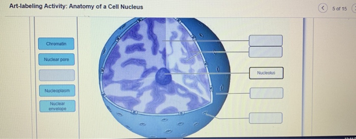
Solved Art-labeling Activity: Anatomy of a Cell Nucleus ② ...
Solved need help labeling these. the first picture is the | Chegg.com This problem has been solved! See the answer. need help labeling these. the first picture is the animal epithelium ,400x ( label the microvilli, cytoplasm, goblet cell with vacuole and nucleus) . the second picture is adipocytes ,400x (label the nucleus , cytoplasm and vacuole )
Carbon Atom Molecular Structure Labels Stock Vector ...
📐identfy the parts of he atom that are labeied in the dagram Label A ... The protons are positively charged, the electrons are negatively charged and the neutrons are neutral that means it has no charge. In the picture, label A shows the nucleus. Electrons has the ability to revolve around the nucleus in a fixed circular orbit. Electron cloud represents the spreading of the electrons energy.
140,562 Labelled cell Images, Stock Photos & Vectors ...
animal cell picture and labels cell animal labeled cells diagram structure typical figure celula imagen 3d biology labels membrane para parts label organelles nucleus discovery. Ch. 1 Lesson 1 Cells (5th) - YouTube . 5th cells. Plants VS Animal Cell Diagram Label Black White | Plant And Animal . pulpbits. 35 Label Animal Cell Quiz - Labels ...
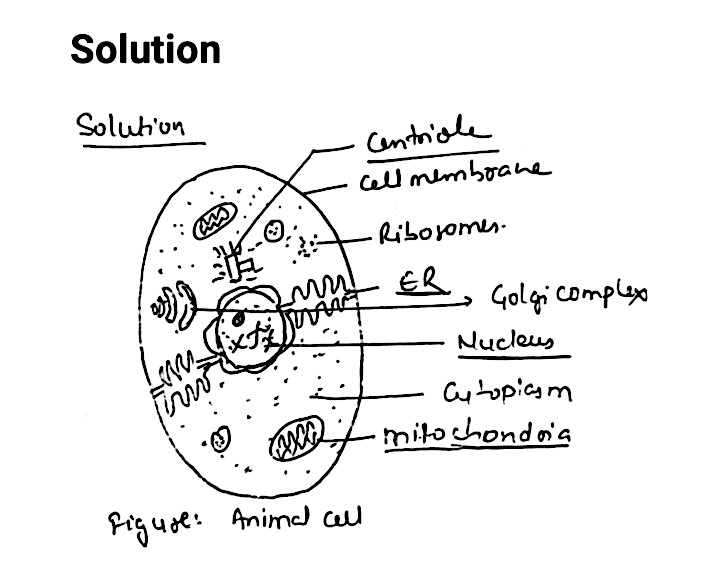
Draw an animal cell and label the cell membrane, nucleus ...
cell picture and labels Animal Cell Picture Without Labels - ClipArt Best . cell without labels animal cliparts computer designs use. Used Black Socks: August 2009 ... cell labeled typical lysosome clipart diagram vector animal illustration drawing nucleus illustrations shutterstock structure royalty prints eps10 cells medical freeart. 33 Plant Cell ...
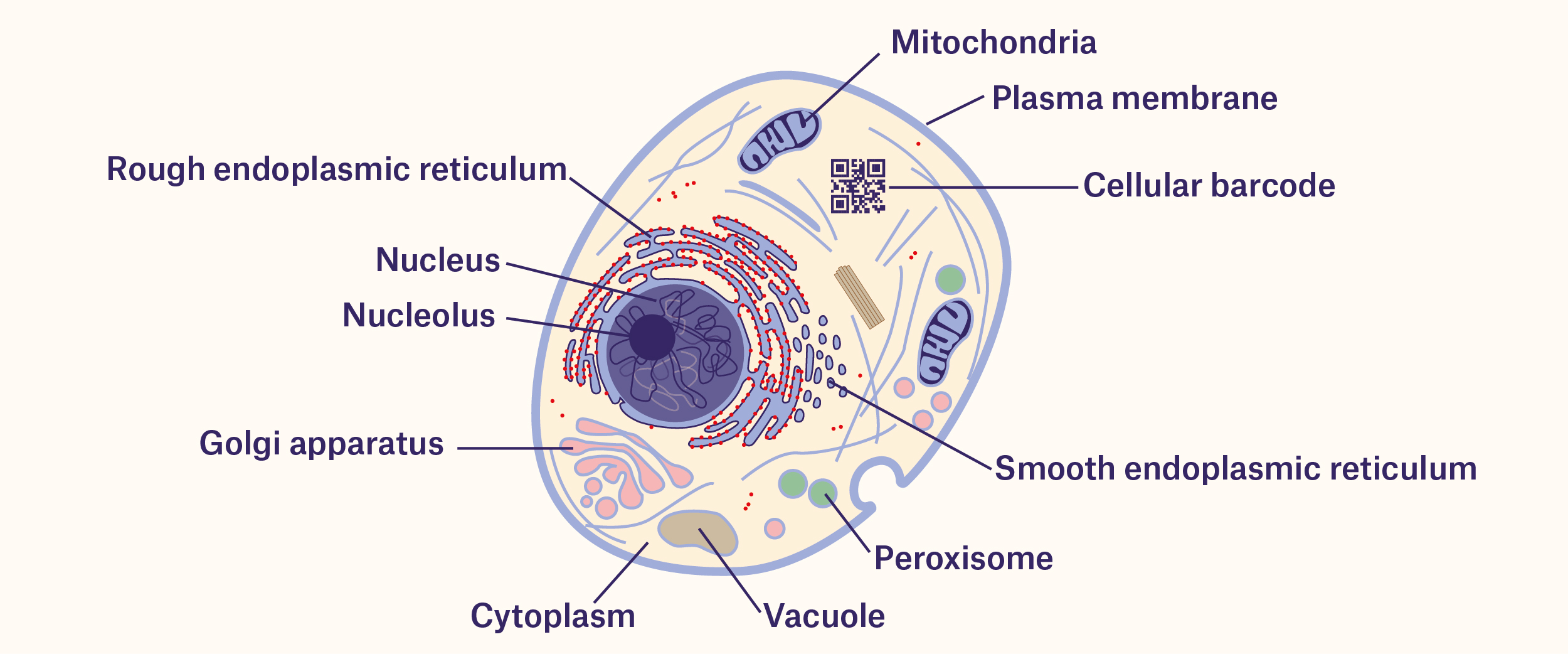
Tracking Cells With Barcodes: Beyond the Label - Labtag Blog
How to draw & label the nucleus - YouTube A beautiful drawing of the nucleus. And it will teach you draw Nucleus very easily. Watch the video and please be kind enough to thumbs up my videos. And I w...
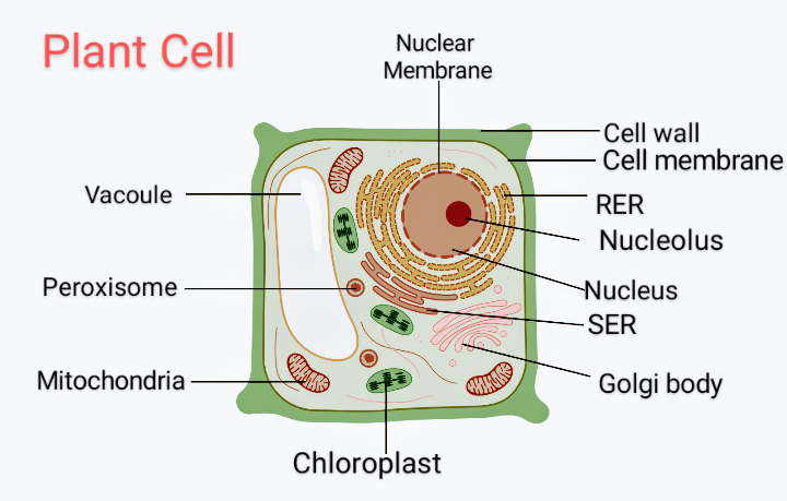
Draw a diagram of a plant cell and label at least eight class ...
How to draw & label the nucleus - Pinterest The space between the inner and outer layers is called Pericardial space, it is filled with pericardial fluid. Pericardium and pericardial fluid protect the heart from physical shocks. Now lets start drawing the diagram. 1.Draw a oblique line from Right to Left 2.Draw a cone shape as shown in… J jessica Heart anatomy drawing Anatomy Drawing

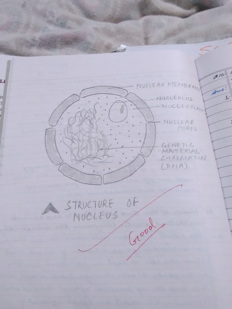
Structure of Nucleus | How to Draw Structure of Nucleus | Nucleus Structure Diagram
What is a Nucleus?- Structure and Function of Nucleus - Byju's The nucleus is a double-membraned organelle that contains the genetic material and other instructions required for cellular processes. It is exclusively found in eukaryotic cells and is also one of the largest organelles. Outline the structure of the Nucleus. A double-membraned organelle known as the nuclear membrane/envelope engirdles the nucleus.
What is a Nucleus?- Structure and Function of Nucleus
Schematic outline of the steps involved in nucleus labeling ...

Label the cell structure. | Study.com

Cell Nucleus - label Diagram | Quizlet

Nucleus - Paige's Cell Project

Blausen 0212 - Nucleus of the cell - English labels | AnatomyTOOL

ROI samples (from weak-label-set) for nucleus label ...
Draw the nucleus and label its parts What is the function ...

How to draw & label the nucleus
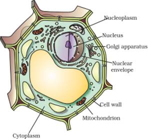
Draw a plant cell and label the parts which (a) determines ...
Spirogyra Cell Anatomy Algae Labeling Cell Stock Vector ...

Draw a well labelled diagram of nucleus.
Draw and label the structure of Nucleus. - Sarthaks eConnect ...

Whole nuclei showing various configurations of satellite ...

The Structure and Functions of a Cell Nucleus Explained ...
Draw a picture of an animal cell. Label the nucleus ...
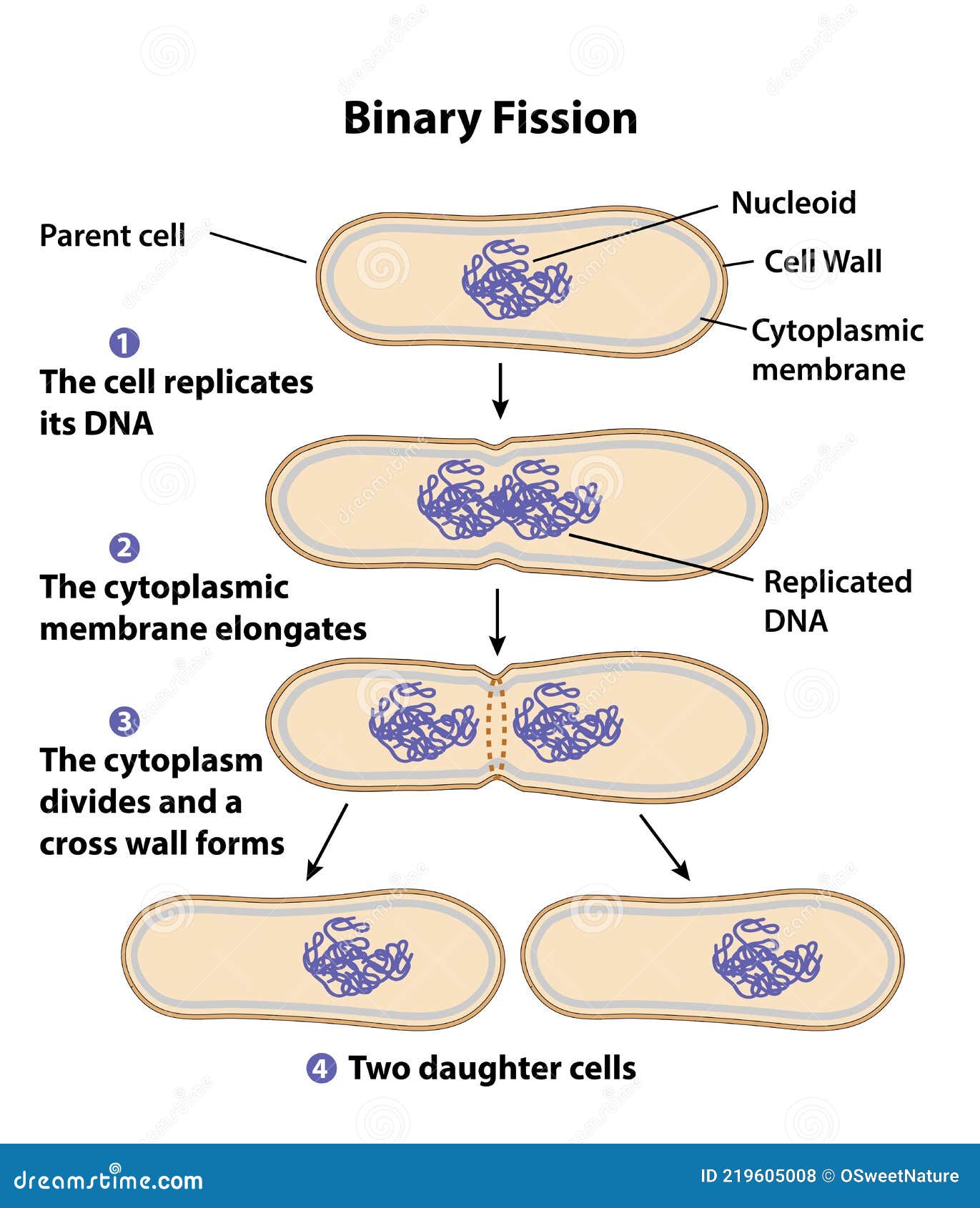
Binary Fission Diagram with Labels Stock Vector ...

cells intro
Cell nucleus hi-res stock photography and images - Alamy
Nucleus Images – Browse 97,718 Stock Photos, Vectors, and ...
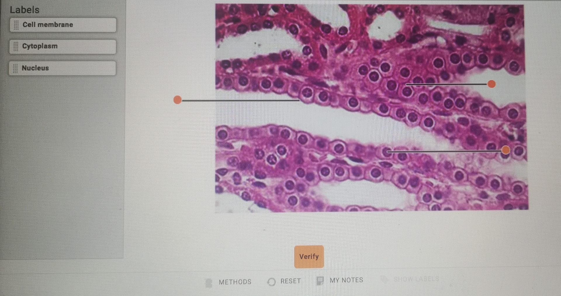
Solved Labels Cell membrane Cytoplasm co Nucleus Verify ...
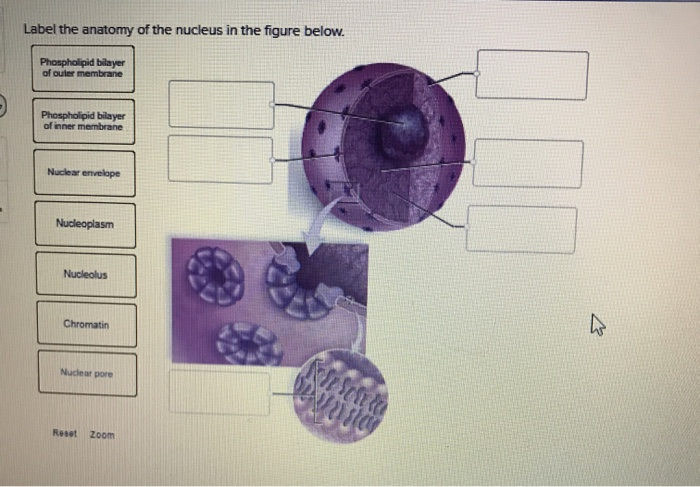
Solved Label the anatomy of the nucleus in the figure below ...
draw a well label diagram of eukaryotic nucleus - Science ...

Pin page

Describe the structure of nucleus.
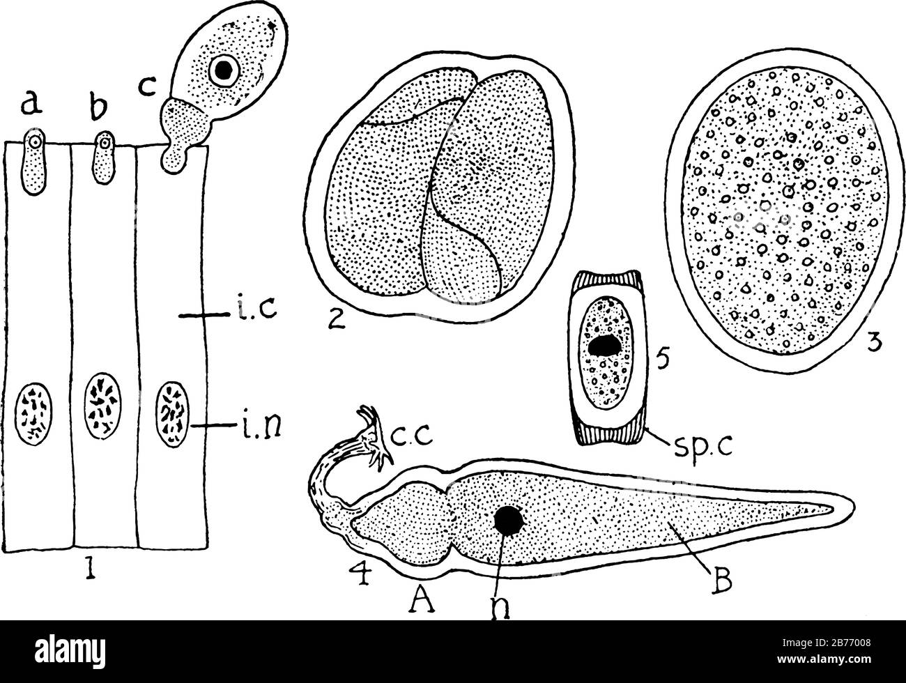
Labels, 1, Young forms (a, b, c) emerging from intestinal ...
A dual-strategy expression screen for candidate connectivity ...

Parts of an atom Label the diagram Atoms
Post a Comment for "41 nucleus picture with labels"