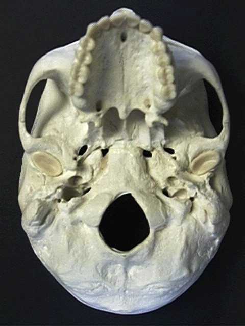
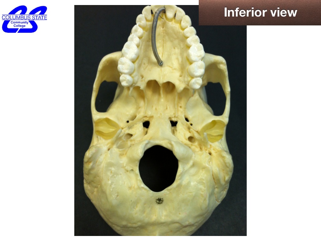
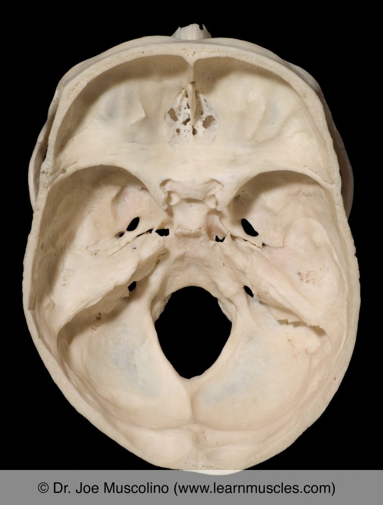
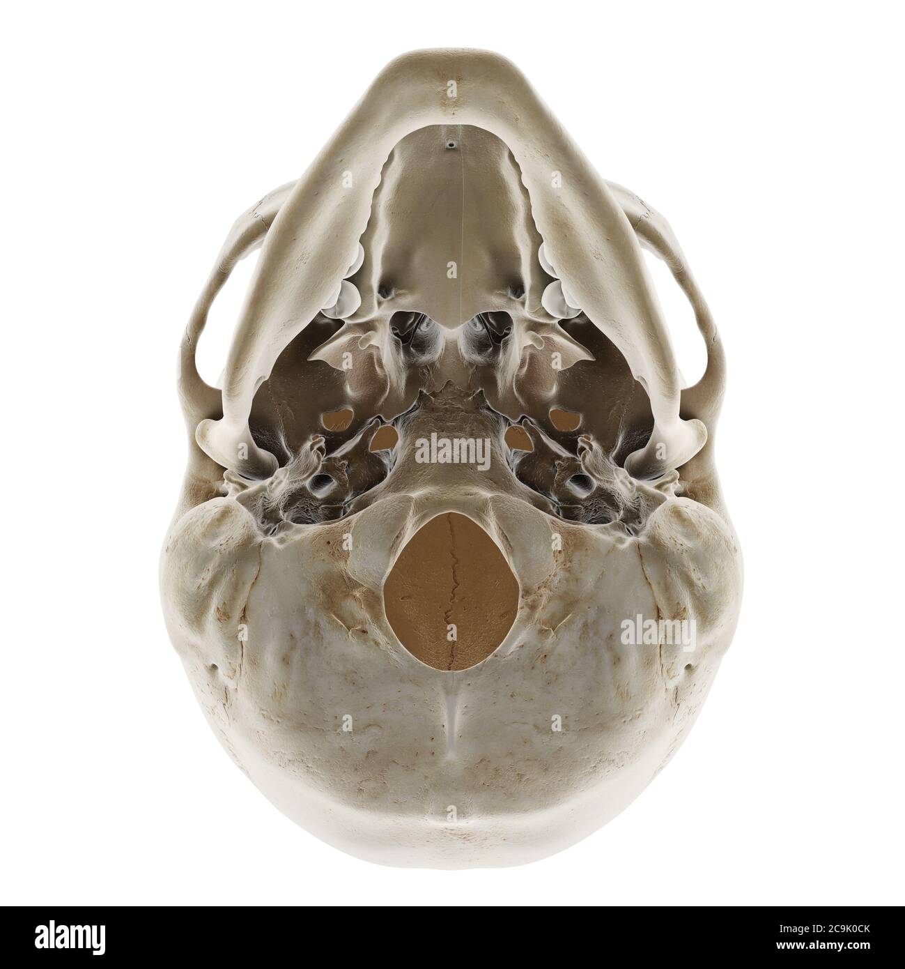
44 inferior view of skull unlabeled
PDF Inferior View of Skull (unlabeled) - SLCC Anatomy Inferior View of Skull (unlabeled) Created Date: 3/18/2015 2:45:00 AM ... Bones and Landmarks seen in the Lateral View of the Skull (Pterion ... Bones and Landmarks Seen in the Inferior View of the Skull Incisive Fossa: Location, Connection, and Transmissions Greater and Lesser Palatine Foramina: Location, Connection, and Transmission Posterior Nasal Spine and Posterior Nasal Aperture
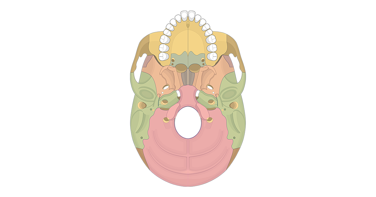
Inferior view of the base of the skull: Anatomy | Kenhub The parietal bones are difficult to visualise from the inferior view of the skull, however they can be seen articulating with the temporal and occipital bones. They form the posterosuperior part of the skull. Clinical points Young children who present with cleft palate have a failure of the two maxillae to unite in the midline.

Inferior view of skull unlabeled
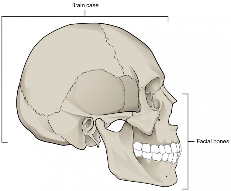
PDF Skull Cranial skeleton (Neurocranium) Skull - 11 Skull: lateral view Frankfurt plane (anatomical position, OrbitoMeatal line): upper margin of ext. acoustic meatus - orbit floor →horizontal superior temporal line; inferior temporal line external acoustic meatus; mastoid process level of ant., mid., post. cranial fossae Unlabeled Skull Diagram The Splanchnocranium is the facial skeleton. The Neurocranium is the braincase. The skull in infants is made up of 45 separate elements but as an adult it is normally made up of 28 elements (including the ear ossicles) (White & Folkens 77). Jul 11, · Printable Eye Diagram Quiz Unlabeled on Diagram Site. Bones of the Skull- INFERIOR VIEW Diagram | Quizlet Bones of the Skull- INFERIOR VIEW + − Flashcards Learn Test Match Created by caseyjc2002 Terms in this set (16) what does point 1 show? The Zygomatic Bone what does point 2 show? The Sphenoid Bone what does point 3 show? The Vomer what does point 4 show? The Occipital Condyle what does point 5 show? The Foramen Magnum what does point 6 show?
Inferior view of skull unlabeled. Success Essays - Assisting students with assignments online Get 24⁄7 customer support help when you place a homework help service order with us. We will guide you on how to place your essay help, proofreading and editing your draft – fixing the grammar, spelling, or formatting of your paper easily and cheaply. A 3D stereotactic atlas of the adult human skull base Figure 1 shows the internal (superior view) and external (inferior view) surfaces of the skull base. ... The images are unlabeled or only partly labeled. Atlas of Skull Base Surgery and Neurotology by Jackler focuses on surgical illustrations with text limited to describe operative techniques. Generally, the textbook materials including print ... Skull - Inferior View - Human Body Help Skull - Inferior View. Quiz yourself with the picture below. Scroll down for the answer key. Videos are at the bottom if you want to watch them first. Key: Incisive fossa. Horizontal plate of the palatine bone. Medial pterygoid plate. Lateral pterygoid plate. posterior view of skull unlabeled - youstalkingmenow Pictures of skulls that are unlabeled and has empty boxes for students to add labels. Our intention is that these Skull Inferior View Worksheet pictures gallery can be a guidance for you deliver you more ideas and of course make you have what you search. This is an online quiz called Sphenoid Bone superior view.
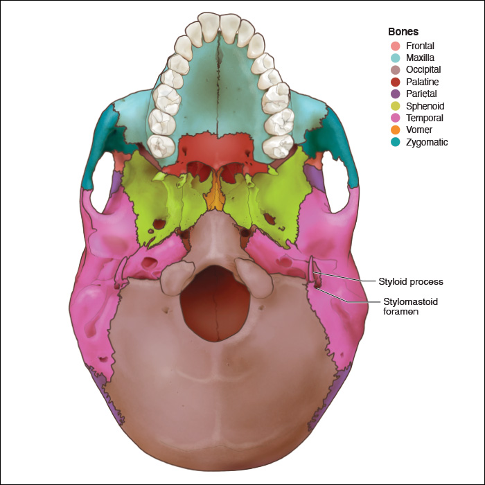
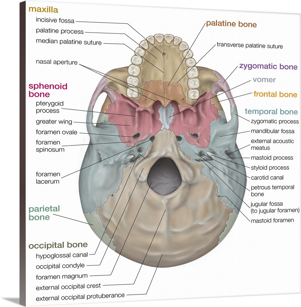
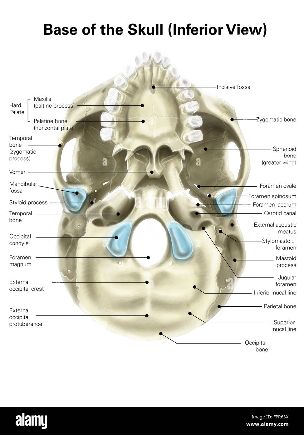
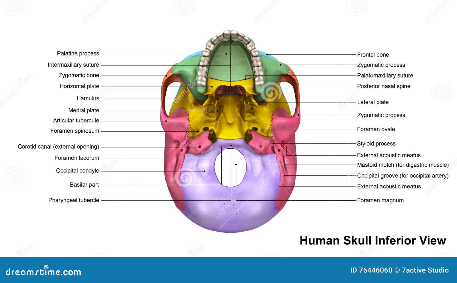
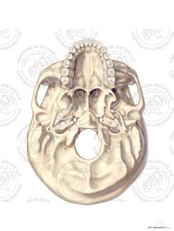
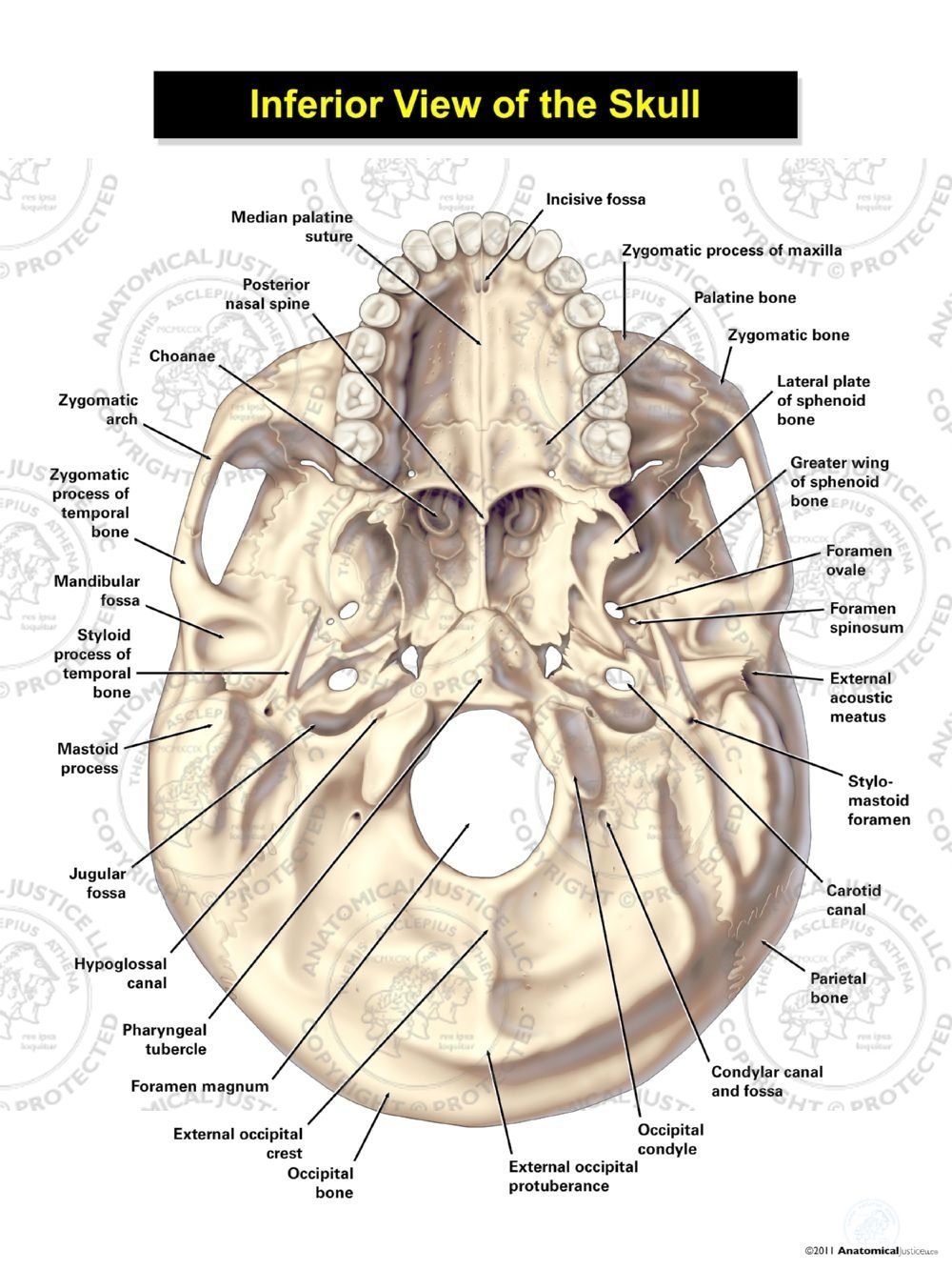
Inferior View of the Skull - Anatomical Justice Inferior View of the Skull. SKU: S04021. This exhibit depicts the anatomy of the inferior skull including: the foramen magnum, occipital condyles, mastoid process, styloid process, mandibular fossa, palatine bone, sphenoid bone, carotid canal, and the jugular fossa. Base Stock Illustration Fee. Hole's Human Anatomy & Physiology | The study of the human body Lateral view of the skull Figure 7.21 Unlabeled; Inferior view of the skull Figure 7.22; Inferior view of the skull Figure 7.22 Unlabeled; The sphenoid bone Figure 7.23A-B; The sphenoid bone Figure 7.23A-B Unlabeled; The ethmoid bone Figure 7.24A-B; The ethmoid bone Figure 7.24A-B Unlabeled; Join LiveJournal Password requirements: 6 to 30 characters long; ASCII characters only (characters found on a standard US keyboard); must contain at least 4 different symbols; The Skull- Inferior view Flashcards | Quizlet Foramen spinosum. Greater wing of sphenoid bone. Hypoglossal canal. Jugular foramen. Mastoid process. Maxilla. Occipital bone. Opening of carotid canal. Temporal bone (inferior aspect of petrous part)
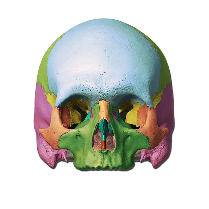
7.2 The Skull - Anatomy and Physiology | OpenStax A better view of the vomer bone is seen when looking into the posterior nasal cavity with an inferior view of the skull, where the vomer forms the full height of the nasal septum. The anterior nasal septum is formed by the septal cartilage, a flexible plate that fills in the gap between the perpendicular plate of the ethmoid and vomer bones ... Skull anatomy: Anterior and lateral views of the skull | Kenhub The bones of the skull that are visible from an anterior and a lateral view are the following: the sphenoid bone (with the greater and the lesser wings) the frontal bone (especially the orbital surface) the zygomatic bone the maxilla the mandible the nasal bones the ethmoid bones the parietal bone and the temporal bone The Skull · Anatomy and Physiology A better view of the vomer bone is seen when looking into the posterior nasal cavity with an inferior view of the skull, where the vomer forms the full height of the nasal septum. The anterior nasal septum is formed by the septal cartilage, a flexible plate that fills in the gap between the perpendicular plate of the ethmoid and vomer bones ... Long Bone Diagram Unlabeled Parts Of A Long Bone. Image of a typical long bone is shown with numbers identifying the various parts, such as the epiphysis. Blank Skull Diagram - Blank Long Bone Diagram Popular Skull diagram unlabeled skull diagram inferior view template information title. Most, but not all, features you are required to know are shown on the following pages.
Skull inferior view Diagram | Quizlet
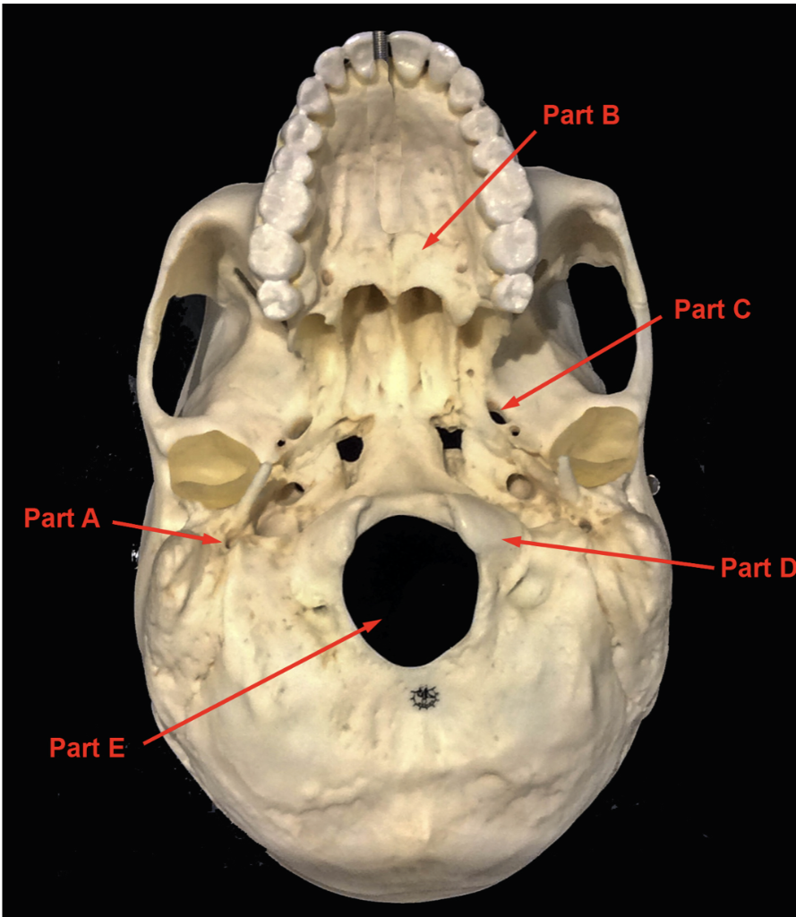
The Skull | Anatomy and Physiology I | | Course Hero On the inferior aspect of the skull, each half of the sphenoid bone forms two thin, vertically oriented bony plates. These are the medial pterygoid plate and lateral pterygoid plate (pterygoid = "wing-shaped"). The right and left medial pterygoid plates form the posterior, lateral walls of the nasal cavity.
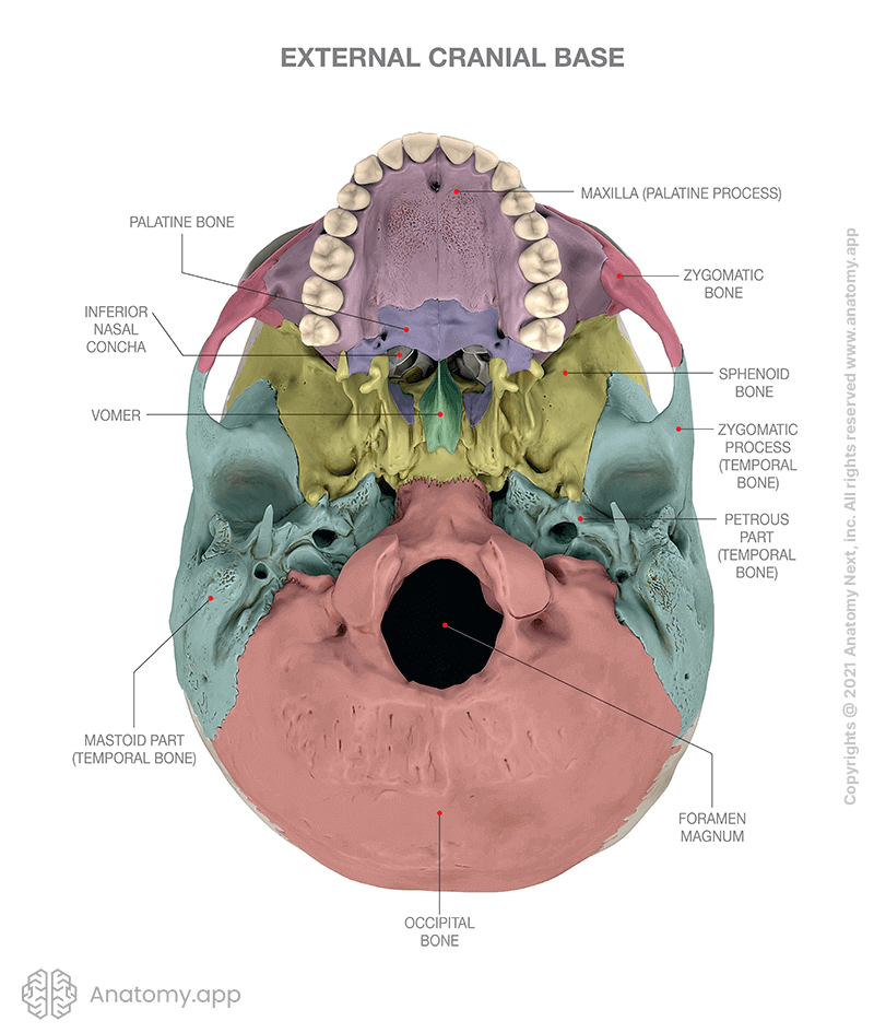
External cranial base | Encyclopedia | Anatomy.app | Learn ...
Solved Diagram (or obtain an unlabeled digital image of) the - Chegg Diagram (or obtain an unlabeled digital image of) the skull in the various orientations/views listed below. Complete the following tasks for Pre-Lab #3. Label all of the components listed in Axial Skeleton Part A, the Skull. Feel free to color or be creative as well. AXIAL SKELETON - PART A - THE SKULL - Sutures. coronal (frontal) suture
Human Skull, Inferior View
Skull Labeling Worksheet Answers Lateral View Of The Bones Of The Skull Unlabeled L1345175778985 Jpg 849 634 Anatomy Bones Human Skull Anatomy Anatomy And Physiology. Skull Labeling Worksheet Image Search Results Worksheets Free Worksheets Anatomy. ... Bones Of The Skull Inferior Anatomy Bones Skull Anatomy Anatomy.
Temporal Bones - Head and Neck Anatomy: Part I – Bony ...
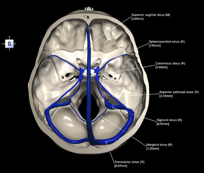
Related image | Anatomy bones, Skull anatomy, Anatomy - Pinterest The detailed view of the inner surface of the cranial base demonstrating openings and connections to other topographical areas of the skull. Anatomy Standard. Topography of the Skull. Nursing Student Tips. Nursing School Studying. Nursing Notes. Medical School. Ear Anatomy. Anatomy Study.
8.2.1: Exterior of the Cranium - Biology LibreTexts
Journal of Neural Engineering - IOPscience Journal of Neural Engineering was created to help scientists, clinicians and engineers to understand, replace, repair and enhance the nervous system.. Transparent peer review is available.
Skull - inferior view. skeletal system Solid-Faced Canvas Print
Skull inferior view Quiz - PurposeGames.com An unregistered player played the game 3 hours ago About this Quiz This is an online quiz called Skull inferior view There is a printable worksheet available for download here so you can take the quiz with pen and paper. From the quiz author Human skull review Your Skills & Rank Total 0 Get started! Today's Rank -- 0 19
Inferior view of the base of the skull: Anatomy | Kenhub
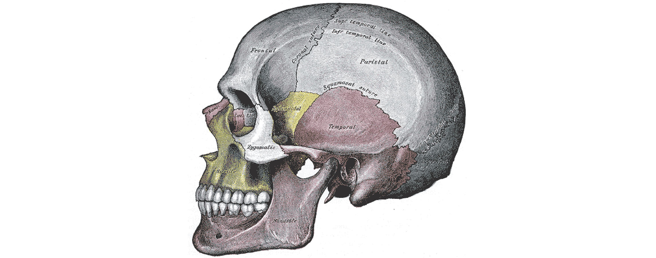
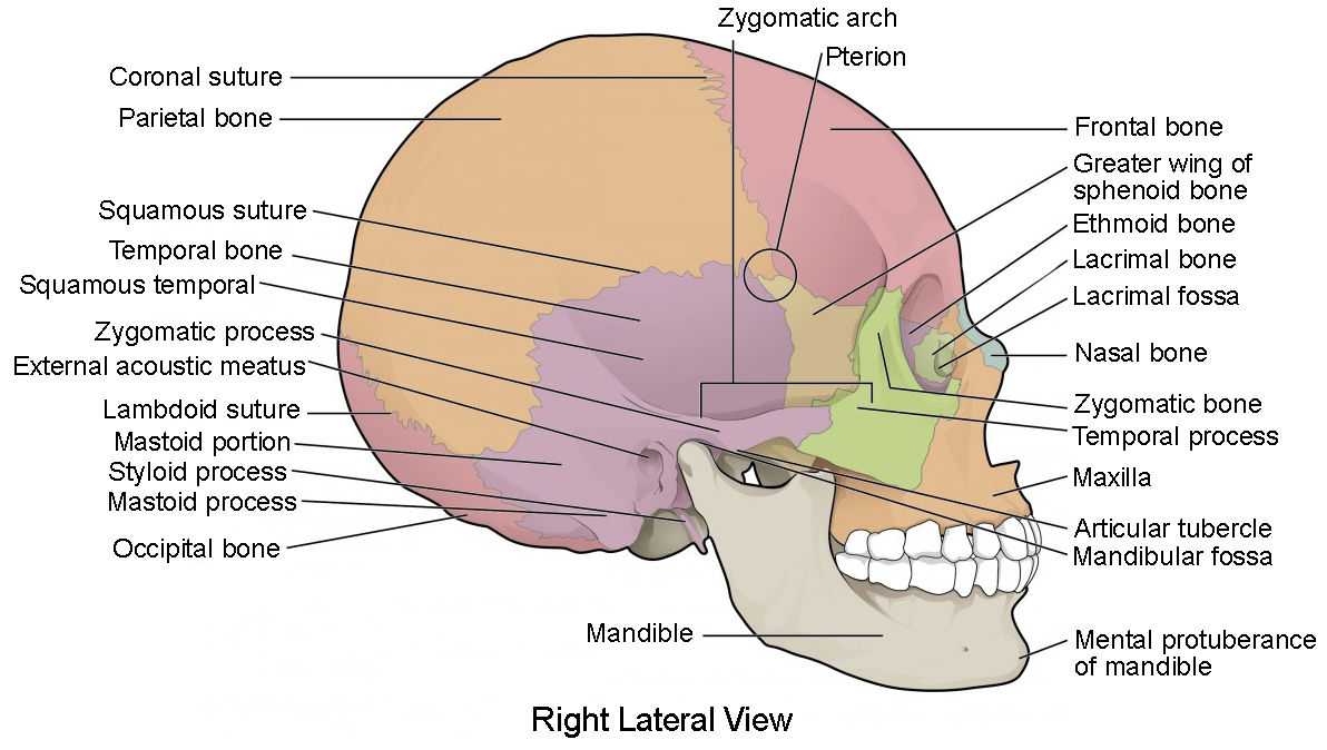
Skull Overview - Head and Neck Anatomy: Part I - Bony Structures ... The other set, while they can be seen on a lateral view of the skull is best understood with a diagram showing the inferior view of the skull (Appendix C). ... The space above the zygomatic arch (This is unlabeled but is where area where the pink and aqua bones meet in Appendix B) is the temporal fossa and below that is found the infratemporal ...

Human Skull: Facial and Cranial Bones Quiz - By kfastic
Skull: Anterior View - Netter Images ID: 14669 Title: Skull: Anterior View Category: Labeled - Flash Cards ID: 61976 Title: Skull: Anterior View Category: Labeled-Cochard Imaging 1E ID: 47728 Title: Skull: Anterior View Category: Labeled-Hansen FC 3E
Inferior view of the skull
Anterior view of Skull Quiz - PurposeGames.com This is an online quiz called Anterior view of Skull There is a printable worksheet available for download here so you can take the quiz with pen and paper. From the quiz author basic parts of the skull from the anterior view This quiz has tags. Click on the tags below to find other quizzes on the same subject. Anatomy anterior Your Skills & Rank
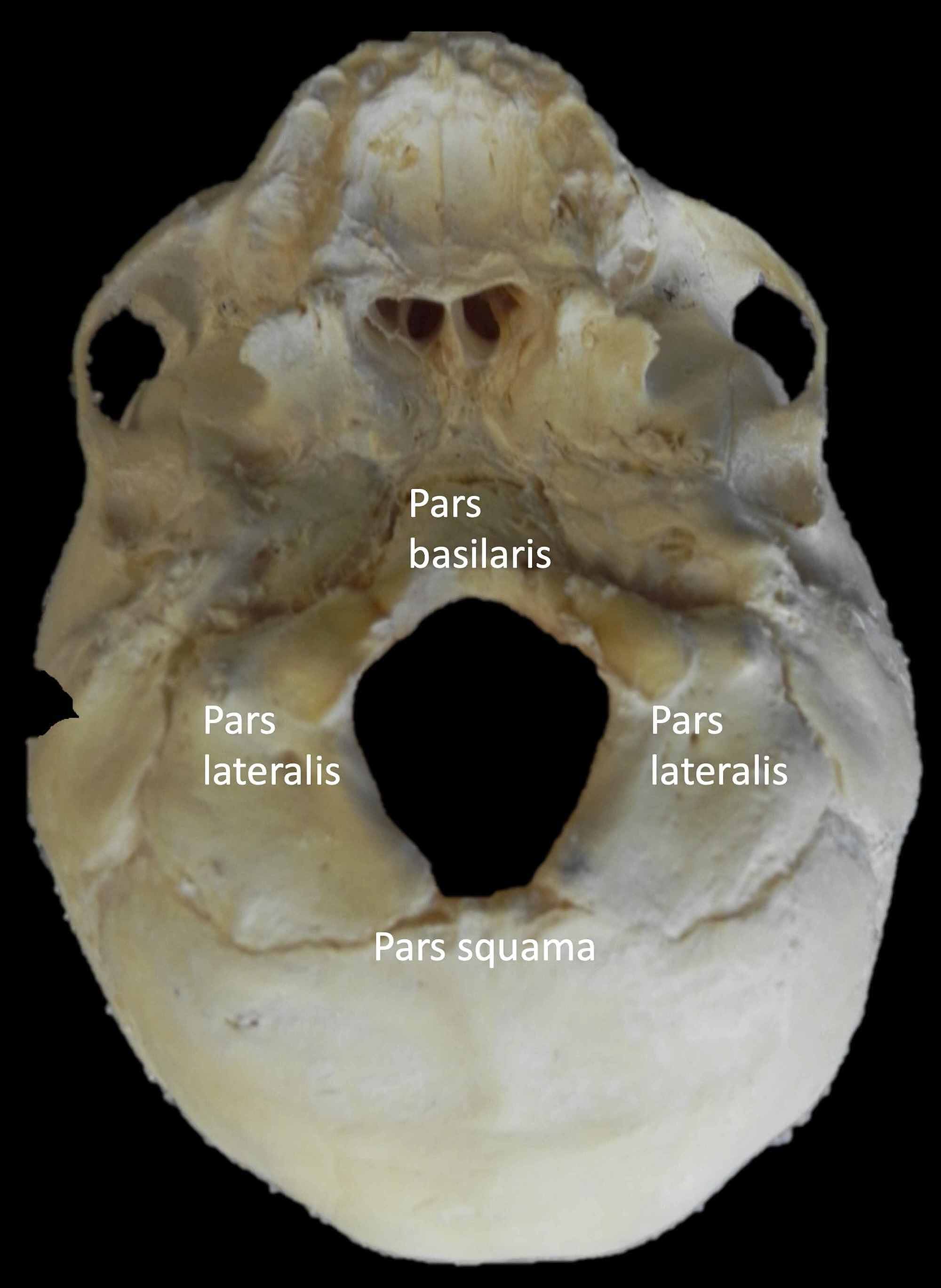
Cureus | Foramen Magnum Variant With Elongation of the ...
The Skull Bones Anatomy - Inferior View | GetBodySmart Let's start with taking a look at the cranial and facial bones from an anterior view before we dive into their markings from an inferior perspective. Facial Bones: Zygomatic bone ( os zygomaticum ). Maxilla bone ( os maxilla ). Palatine bone ( os palatinum ). Learn skull anatomy faster with these interactive skull bones quizzes and worksheets.
9 The Axial Skeleton
TransparencyList11e.doc - List of Transparencies TO... 167. Lateral view of the skull, unlabeled Figure 7.12 168. Inferior view of the skull Figure 7.13 169. Inferior view of the skull, unlabeled Figure 7.13 170. Floor of the cranial cavity Figure 7.14 171. Floor of the cranial cavity, unlabeled Figure 7.14 172. Sagittal section of the skull Figure 7.15 173. Sagittal section of the skull, unlabeled Figure 7.15 174. ...

Colored Base Of Human Skull Inferior View With Labels High ...
Junqueira's Basic Histology Text and Atlas, 14th Edition Download Free PDF View PDF. SECTION-1: GENERAL PROPERTIES. HD Dynasty. Download Free PDF View PDF. HISTOLOGY FULL-TEXT. Ahmed EL-Masry. Download Free PDF View PDF ...
Learn skull anatomy with skull bone quizzes and diagrams | Kenhub
Foramina of Cranial Base: Inferior View - Netter Images Foramina of Cranial Base: Inferior View Variant Image ID: 8790 Add to Lightbox. Save to Lightbox. Email this page; Link this page ; Print; Please ... Skull: Basal View Category: Labeled-Weber. This Illustration was published in. Atlas de Anatomia Humana Author: Frank H. Netter, MD Chapter: Cabeça e Pescoço Page: 10.
7.2 The Skull - Anatomy and Physiology 2e | OpenStax
Bacterial sensing via neuronal Nod2 regulates appetite and ... A small craniotomy was performed and the viral solution was then injected into DMH and/or ARC through a glass micropipette attached to a nanoinjector system (Nanoject III; 100 nl per site; 1 nl/s) using stereotaxic coordinates (–1.7 mm anterior from bregma, 0.3 mm lateral, and at a depth of 5 mm for DMH and 5.9 mm for ARC from the skull ...
Base of human skull, inferior view, with labels Stock Photo ...
Inferior Skull Bones Quiz | GetBodySmart Inferior Skull Bones Quiz. Author: Scott A. Sheffield MS. Last update: Sep 8th, 2022. Learn anatomy faster and. remember everything you learn.
9 The Axial Skeleton
Labeled+Inferior+View+of+the+Superficial+Skull.PNG 949×596 pixels ... Aug 5, 2014 - Labeled+Inferior+View+of+the+Superficial+Skull.PNG 949×596 pixels. Aug 5, 2014 - Labeled+Inferior+View+of+the+Superficial+Skull.PNG 949×596 pixels. ... SmartDraw includes Anatomy examples like this Vertebra Unlabeled template that you can easily edit and customize in minutes. Belinda Curtis. Education. Skull Anatomy. Head Anatomy.
Skull ○Cranial skeleton (Neurocranium) ○Facial skeleton ...
Bones of the Skull- INFERIOR VIEW Diagram | Quizlet Bones of the Skull- INFERIOR VIEW + − Flashcards Learn Test Match Created by caseyjc2002 Terms in this set (16) what does point 1 show? The Zygomatic Bone what does point 2 show? The Sphenoid Bone what does point 3 show? The Vomer what does point 4 show? The Occipital Condyle what does point 5 show? The Foramen Magnum what does point 6 show?
Skull Inferior View Stock Illustrations – 60 Skull Inferior ...
Unlabeled Skull Diagram The Splanchnocranium is the facial skeleton. The Neurocranium is the braincase. The skull in infants is made up of 45 separate elements but as an adult it is normally made up of 28 elements (including the ear ossicles) (White & Folkens 77). Jul 11, · Printable Eye Diagram Quiz Unlabeled on Diagram Site.
Skull Labeling Quiz
PDF Skull Cranial skeleton (Neurocranium) Skull - 11 Skull: lateral view Frankfurt plane (anatomical position, OrbitoMeatal line): upper margin of ext. acoustic meatus - orbit floor →horizontal superior temporal line; inferior temporal line external acoustic meatus; mastoid process level of ant., mid., post. cranial fossae
Inferior View of Skull | Science, anatomy | ShowMe

The skull

Unit 4 - The Skeletal System - The Skull (Inferior View ...
A 3D stereotactic atlas of the adult human skull base | Brain ...

Skull - Inferior View (Photos) - Gross Anatomy Flashcards ...
Skull - Learn Muscles

Cranial Bones (Inferior View) Quiz

Skull Inferior view stock illustration. Illustration of face ...

Cranial and Facial Bones Inferior View Diagram | Quizlet
The Skull | Anatomy and Physiology I | | Course Hero

Human Skull Anatomy Inferior View (Illustrations) – Human Bio ...
Axial Skeleton

Skull - Inferior View - Gross Anatomy Flashcards | Draw it to ...

Bones and Features of the Skull – David Fankhauser

The Skull Bones Anatomy - Inferior View | GetBodySmart
The Skull | Anatomy and Physiology I | | Course Hero

SKULL - OPENINGS (Inferior View) Quiz
Solved Inferior view of the skull. Part A: What is the name ...
Foramen magnum hi-res stock photography and images - Alamy

Inferior View of Skull Quiz (KNES 259)
The Skull Bones Anatomy - Inferior View | GetBodySmart
Inferior View of the Skull

Occipital bone labeled: anatomy & landmarks | GetBodySmart
Inferior View of the Skull

Skeleton-labeling-worksheet & Blank Skeleton Diagram To Label ...
Post a Comment for "44 inferior view of skull unlabeled"