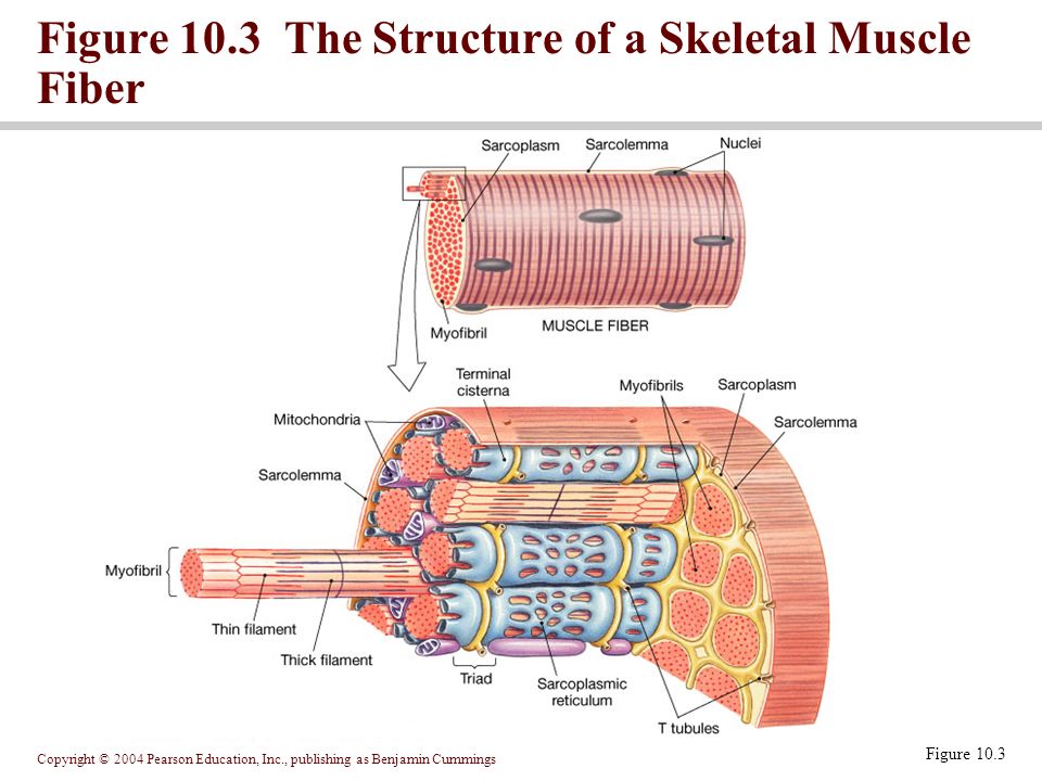
38 label the structures of a skeletal muscle fiber.
Label structure of skeletal muscle Diagram | Quizlet Label structure of skeletal muscle Diagram | Quizlet Label structure of skeletal muscle 4.0 (5 reviews) + − Learn Test Match Created by danielaaaa04 Terms in this set (8) myofibrils ... sarcoplasmis reticulum ... sarcolemma ... epimysium ... perimysium ... endomysium ... fascicle ... muscle fiber ... Correctly Label The Following Parts Of A Skeletal Muscle Fiber The sarcomeres of a skeletal muscle fiber have a striated appearance and are located between the myofibrils. The sarcoplasm is a specialized structure that stores calcium ions and glycine. These three components work together to make up the skeletal muscle fiber. You should be able to label each of these parts accurately if you want to ...
ANSWER THIS NOW!!!! 40 points!!! Drag each label to the correct ... Answer: 1. Muscle: soft, contractile tissue important to produce force and motion in animals. 2. Fascicle: multiple bundles of skeletal muscle fibres which is surrounded by a type of connective tissue called perimysium. 3. Muscle fibres: bundles of cylindrical organelles myofibrils formed by the fusion of myoblasts via myogenesis process. 4.

Label the structures of a skeletal muscle fiber.
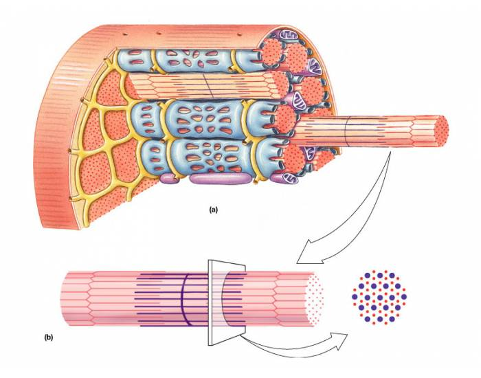
Cells | Free Full-Text | In Adult Skeletal Muscles, the Co-Receptors of ... Canonical Wnt signaling is involved in skeletal muscle cell biology. The exact way in which this pathway exerts its contribution to myogenesis or neuromuscular junctions (NMJ) is a matter of debate. Next to the common co-receptors of canonical Wnt signaling, Lrp5 and Lrp6, the receptor tyrosine kinase MuSK was reported to bind at NMJs WNT glycoproteins by its extracellular cysteine-rich domain. 9.2A: Skeletal Muscle Fibers - Medicine LibreTexts Skeletal muscles are composed of striated subunits called sarcomeres, which are composed of the myofilaments actin and myosin. Learning Objectives Outline the structure of a skeletal muscle fiber Key Points Muscles are composed of long bundles of myocytes or muscle fibers. Myocytes contain thousands of myofibrils. Art-labeling Activity: The Structure of a Skeletal Muscle Fiber Art-labeling Activity: The Structure of a Skeletal Muscle Fiber Diagram | Quizlet Art-labeling Activity: The Structure of a Skeletal Muscle Fiber + − Learn Test Match Created by BabeRuthless0504 Terms in this set (2) Art-labeling Activity: The Structure of a Skeletal Muscle Fiber ... Art-labeling Activity: The Structure of a Skeletal Muscle Fiber
Label the structures of a skeletal muscle fiber.. Structure of Skeletal Muscle | SEER Training Each organ or muscle consists of skeletal muscle tissue, connective tissue, nerve tissue, and blood or vascular tissue. Skeletal muscles vary considerably in size, shape, and arrangement of fibers. They range from extremely tiny strands such as the stapedium muscle of the middle ear to large masses such as the muscles of the thigh. Skeletal Muscle - Anatomy & Physiology - University of Hawaiʻi Muscles attach to bones directly or through tendons or aponeuroses. Skeletal muscles maintain posture, stabilize bones and joints, control internal movement, and generate heat. Skeletal muscle fibers are long, multinucleated cells. The membrane of the cell is the sarcolemma; the cytoplasm of the cell is the sarcoplasm. Muscle Fibers: Anatomy, Function, and More - Healthline Skeletal muscle fibers are classified into two types: type 1 and type 2. Type 2 is further broken down into subtypes. Type 1. These fibers utilize oxygen to generate energy for movement. Type... map.aeonbank.co.jp › aeonbankトップ | 店舗・ATM検索|イオン銀行 全国に設置しているイオン銀行atmや店舗を現在地や駅名などのさまざまな方法で検索できます。イオン銀行のキャッシュカードなら、イオン銀行atmで24時間365日手数料無料。
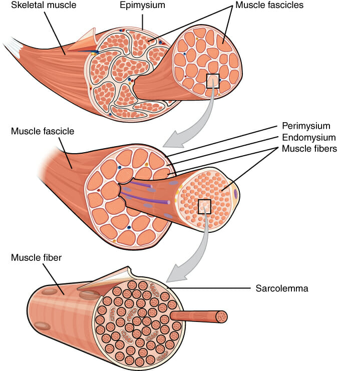
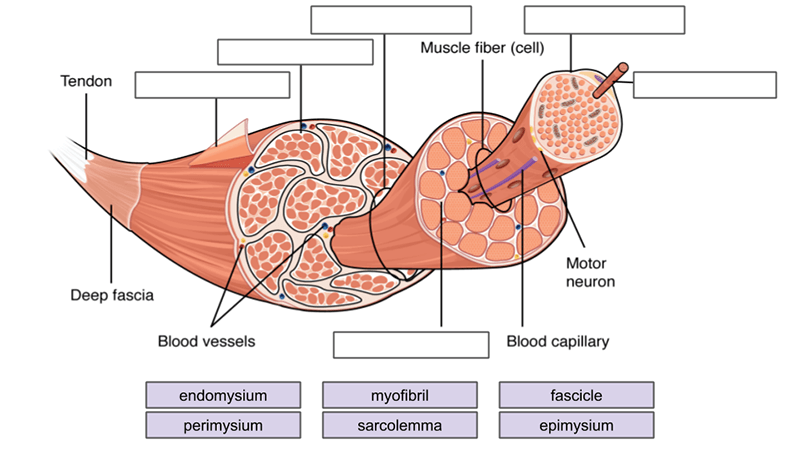
Skeletal Muscle | Anatomy and Physiology I - Lumen Learning These tissues include the skeletal muscle fibers, blood vessels, nerve fibers, and connective tissue. Each skeletal muscle has three layers of connective tissue (called "mysia") that enclose it and provide structure to the muscle as a whole, and also compartmentalize the muscle fibers within the muscle (Figure 1). Microscopic Structure Of Skeleton Muscles | Anatomy Notes In this topic, we will discuss the features of skeleton muscles and the microscopic structure of skeletal muscles in detail. Skeletal muscles are striated and voluntary. It is the most common muscle tissue. It consists of long, parallel multinucleate cells bundled together by collagenous sheaths and through this regular organization allow the ... Structure of Skeletal Muscle - CliffsNotes The sarcolemma, or plasma membrane of the muscle cell, is highly invaginated by transverse tubules (T tubes) that permeate the cell. The sarcoplasm, or cytoplasm of the muscle cell, contains calcium‐storing sarcoplasmic reticulum, the specialized endoplasmic reticulum of a muscle cell. Striated muscle cells are multinucleated. Skeletal Muscle Fiber Labeling Flashcards | Quizlet Skeletal Muscle Fiber Labeling Flashcards | Quizlet Skeletal Muscle Fiber Labeling Term 1 / 16 sarcolemma Click the card to flip 👆 Definition 1 / 16 ... Click the card to flip 👆 Flashcards Learn Test Match Created by gracemersch Terms in this set (16) sarcolemma mitochondrion myofibril what is this whole element? dark a band light i band nucleus
microscopic anatomy of a skeletal muscle fiber Quiz microscopic anatomy of a skeletal muscle fiber — Quiz Information. This is an online quiz called microscopic anatomy of a skeletal muscle fiber. There is a printable worksheet available for download here so you can take the quiz with pen and paper. Muscle Fiber Anatomy Quiz - PurposeGames.com Skeletal muscle fiber model. Science. Creator. cwalsh2. Quiz Type. Image Quiz. Value. 15 points. Likes. 66. Played. 39,233 times. Printable Worksheet. Play Now. Add to playlist. ... This is an online quiz called Muscle Fiber Anatomy. There is a printable worksheet available for download here so you can take the quiz with pen and paper. Skeletal Muscle: What Is It, Function, Location & Anatomy Skeletal muscles comprise 30 to 40% of your total body mass. They're the muscles that connect to your bones and allow you to perform a wide range of movements and functions. Skeletal muscles are voluntary, meaning you control how and when they work. Appointments 216.444.2606. Appointments & Locations. en.wikipedia.org › wiki › Vitamin_B12Vitamin B12 - Wikipedia Vitamin B 12 deficiency can potentially cause severe and irreversible damage, especially to the brain and nervous system. At levels only slightly lower than normal, a range of symptoms such as feeling tired, weak, feeling like one may faint, dizziness, breathlessness, headaches, mouth ulcers, upset stomach, decreased appetite, difficulty walking (staggering balance problems), muscle weakness ...
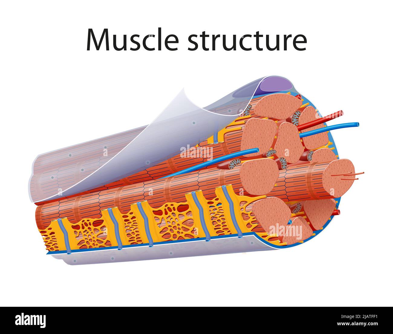
Structure of muscle anatomy. Epimysium covers each muscle ...
10.2 Skeletal Muscle - Anatomy & Physiology These tissues include the skeletal muscle fibers, blood vessels, nerve fibers, and connective tissue. Each skeletal muscle has three layers of connective tissue (called mysia) that enclose it, provide structure to the muscle, and compartmentalize the muscle fibers within the muscle ( Figure 10.2.1 ).

11.2 Muscles and Movement | BioNinja
Solved Muscle Cell Label the structures of a skeletal muscle - Chegg Muscle Cell Label the structures of a skeletal muscle fiber. Nucleus Myofibril Sarcolemma Sarcoplasmic reticulum Openings into T tubules < Prev 3 of 15 !!! Next > Thinkinys - How to write a boty The Good Cre. Dob C ommunicatio pdf Communication.pdf Question: Muscle Cell Label the structures of a skeletal muscle fiber.
Muscle Tissue. - ppt download
Skeletal Muscle Histology Slide Identification and Labeled Diagram ... Please try to find out these structures from the skeletal muscle slide labeled images. #1. Longitudinal section of skeletal muscle #2. Cross-section of skeletal muscle #3. Skeletal muscle fibers of the longitudinal section #3. The nucleus of skeletal muscle fibers in longitudinal and cross-section #4. Cross striations of skeletal muscles #5.

Week 6: Muscle Physiology Flashcards | Quizlet
Skeletal Muscle Tissue Anatomy and Structure - Registered Nurse RN Each skeletal muscle is considered an organ, and it's made up of connective tissue layers, muscle fibers, blood vessels, and nerves. Skeletal muscles attach to the bones through tendons or through a direct attachment. As you look at this muscle diagram, you'll notice an outer layer of connective tissue called epimysium.

Skeletal Muscle | Anatomy and Physiology I
Label the Skeletal Muscle Fiber Quiz - PurposeGames.com Label the Skeletal Muscle Fiber — Quiz Information. This is an online quiz called Label the Skeletal Muscle Fiber. There is a printable worksheet available for download here so you can take the quiz with pen and paper.. From the quiz author

A) Illustration of skeletal muscle structure copied with ...
structure of skeletal muscle fiber Flashcards | Quizlet muscle cell Myofibrils Microscopic protein filaments that make up muscle cells. Myofilaments actin and myosin Epimysium covers the entire skeletal muscle Perimysium The connective tissue that surrounds fascicles. Endomysium Surrounds individual muscle fibers Sarcolemma plasma membrane of a muscle fiber Sarcoplasm cytoplasm of a muscle cell

Label the following in a diagram of a skeletal muscle fiber ...
Skeletal Muscle Fiber | Types, Characteristics & Anatomy - Video ... Skeletal muscles display a nested organization where each structure is composed of individual smaller structures. For instance, each muscle is composed of bundles called fascicles....

Skeletal Muscle Tissue Quiz for Anatomy
Structures of the Skeletal Muscle Fiber Flashcards | Quizlet Muscle Cell or Muscle Fiber or Myofiber -Muscle cells are long, cylindrical & multinucleated -Sarcolemma = muscle cell membrane -Sarcoplasm filled with tiny threads called myofibrils & myoglobin (red-colored, oxygen-binding protein) Transverse Tubules -T (transverse) tubules are invaginations of the sarcolemma into the center of the cell

Study Guide Flashcards | Quizlet
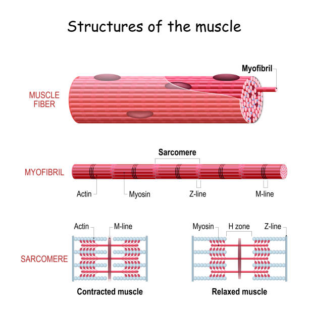
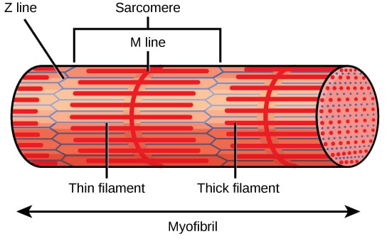
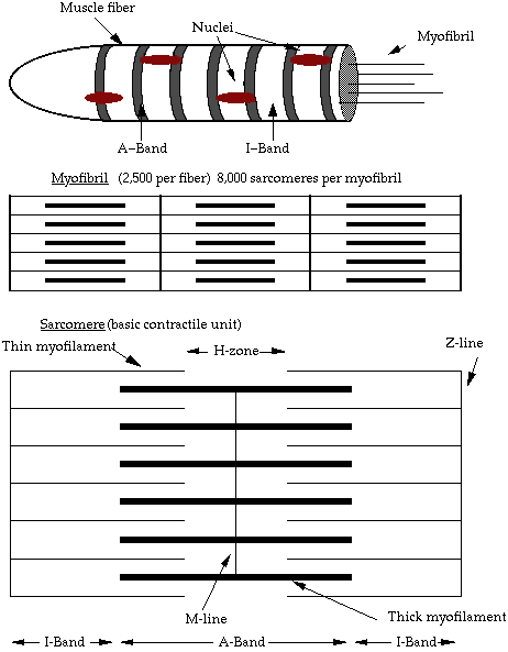
Skeletal Muscle Fiber Structure and Function - Open Textbooks for Hong Kong The striated appearance of skeletal muscle tissue is a result of repeating bands of the proteins actin and myosin that occur along the length of myofibrils. Myofibrils are composed of smaller structures called myofilaments. There are two main types of myofilaments: thick filaments and thin filaments.
3,457 Muscle Cells Stock Photos, Pictures & Royalty-Free ...
Skeletal muscle fibers: arrangement and diagram | GetBodySmart Skeletal muscle fibers are located inside muscles, where they are organized into bundles called fascicles (= fasciculi). 1 2 3 The epimysium is the connective tissue layer that covers the outer surface of the muscle. 1 2 Surrounding and holding together each fascicle is a layer of connective tissue known as perimysium.
Skeletal muscle - Structure - Contraction - TeachMePhysiology
Skeletal Muscle Labeling | Biology Quiz - Quizizz Q. Region where a motor neuron comes in close contact with a muscle cell. answer choices. neurotransmitter. muscular dystrophy. muscle tension. neuromuscular junction. Question 29. 30 seconds. Q. Skeletal Muscle contraction is initiated when the ________ sends a message to the muscle cell.

6,938 Skeletal Muscles Graphic Images, Stock Photos & Vectors ...
Art-labeling Activity: The Structure of a Skeletal Muscle Fiber Art-labeling Activity: The Structure of a Skeletal Muscle Fiber Diagram | Quizlet Art-labeling Activity: The Structure of a Skeletal Muscle Fiber + − Learn Test Match Created by BabeRuthless0504 Terms in this set (2) Art-labeling Activity: The Structure of a Skeletal Muscle Fiber ... Art-labeling Activity: The Structure of a Skeletal Muscle Fiber
Solved] 4. Label the levels of skeletal muscle structure in ...
9.2A: Skeletal Muscle Fibers - Medicine LibreTexts Skeletal muscles are composed of striated subunits called sarcomeres, which are composed of the myofilaments actin and myosin. Learning Objectives Outline the structure of a skeletal muscle fiber Key Points Muscles are composed of long bundles of myocytes or muscle fibers. Myocytes contain thousands of myofibrils.

47.154 Muscle organ Gambar, Foto Stok & Vektor | Shutterstock
Cells | Free Full-Text | In Adult Skeletal Muscles, the Co-Receptors of ... Canonical Wnt signaling is involved in skeletal muscle cell biology. The exact way in which this pathway exerts its contribution to myogenesis or neuromuscular junctions (NMJ) is a matter of debate. Next to the common co-receptors of canonical Wnt signaling, Lrp5 and Lrp6, the receptor tyrosine kinase MuSK was reported to bind at NMJs WNT glycoproteins by its extracellular cysteine-rich domain.
Muscle - Definition, Function, Types and Structure | Biology ...
Muscles Labeling

Neurolemmocyte On Skeletal Muscle Model - Human Anatomy ...

Draw a neuromuscular junction and label every structure ...
Solved] •Label all the parts of the follow diagrams. Muscle ...

Lab Muscle Quiz Flashcards | Quizlet
Muscle Fiber Anatomy Quiz
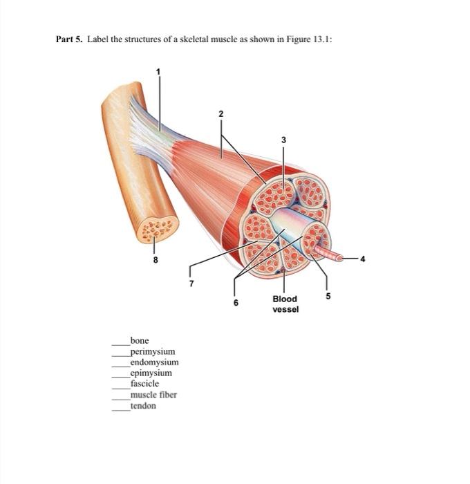
Solved Part 5. Label the structures of a skeletal muscle as ...

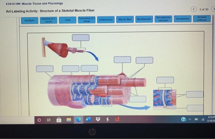
Art-labeling Activity: The Structure of a Skeletal Muscle ...
Solved

The Structure of Skeletal Muscle (6.3.2) | AQA A Level ...
Skeletal muscle hi-res stock photography and images - Alamy

Lab Muscle Quiz Flashcards | Quizlet
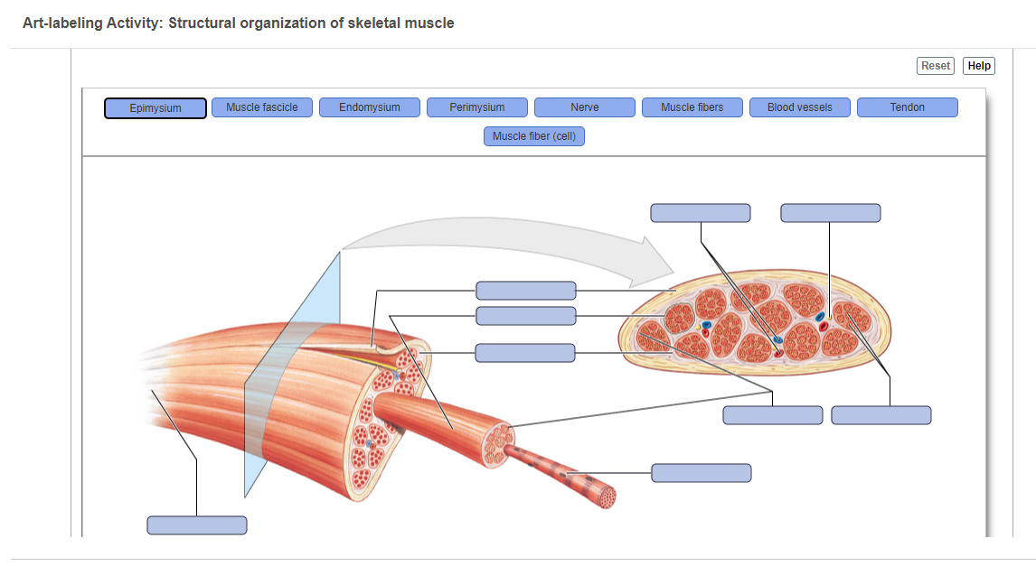
Answered: Art-labeling Activity: Structural… | bartleby
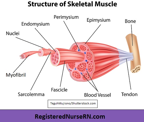
Skeletal Muscle Tissue Anatomy and Structure

muscles-color-and-label-2014-1hwnl96.pdf - Name: Muscles ...

Solved Muscle Cell Label the structures of a skeletal muscle ...
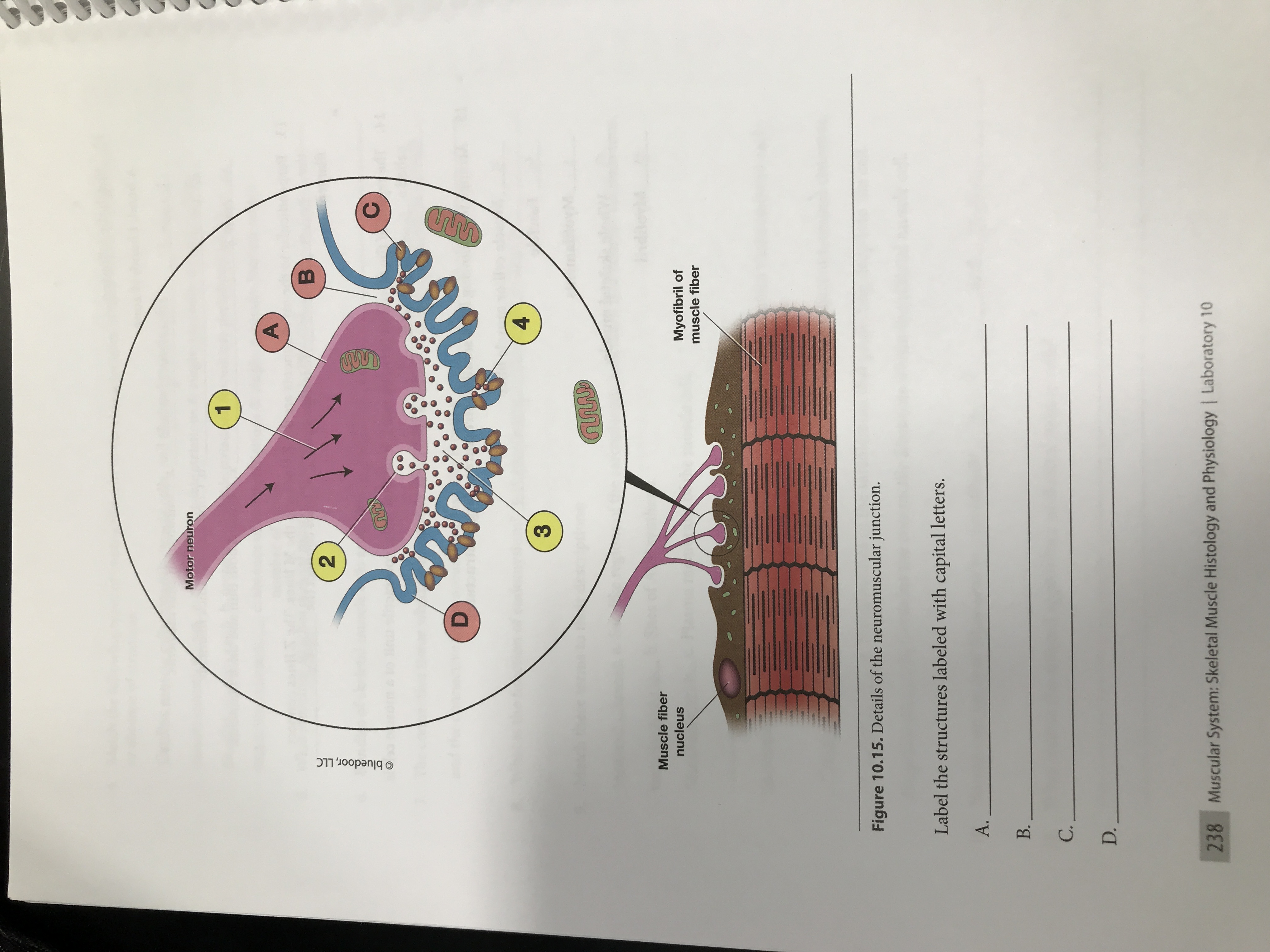
Answered: Motor neuron 4 Muscle fiber nucleus… | bartleby

Organisation d'une fibre musculaire. Les fibres musculaires ...

ANSWER THIS NOW!!!! 40 points!!! Drag each label to the ...
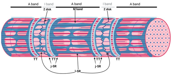
Biomolecules | Free Full-Text | The Sarcoplasmic Reticulum of ...

The structure and internal organization of a skeletal muscle ...

A&P 1- CHAPTER 9 MASTERING ASSIGNMENTS Flashcards | Quizlet
Structure and Composition of Muscle - Meat Science
Post a Comment for "38 label the structures of a skeletal muscle fiber."